
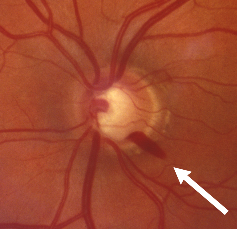
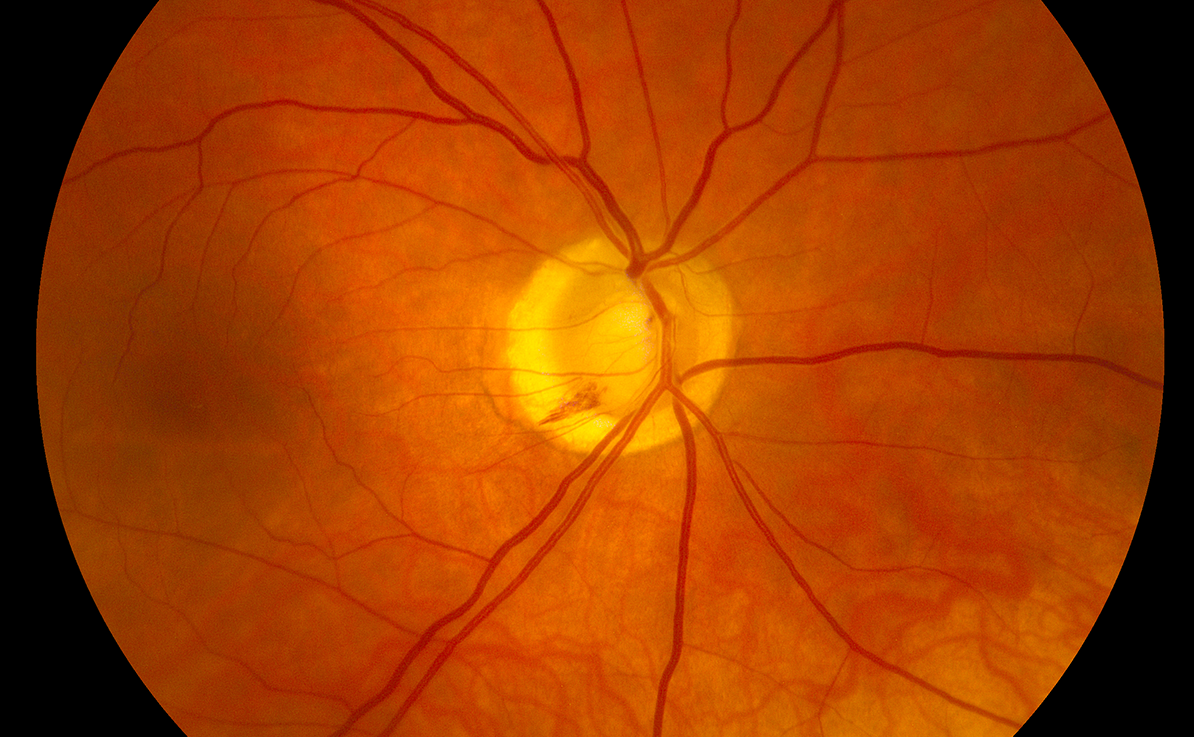
Optic disc hemorrhage is a small, flame-shaped or splinter hemorrhage at or near the optic disc margin, and in glaucoma it is also called a Drance hemorrhage.
It is a well-recognized risk factor for the development and progression of glaucoma.
Finding one should lead to a review of the patient’s pressure, disc, and visual field, and it should not be dismissed as an incidental finding.
What Is an Optic Disc Hemorrhage?
The hemorrhage lies in the superficial nerve fiber layer or at the border of the neuroretinal rim, and it is usually oriented radially along the nerve fibers.
It appears red and sharply demarcated, and it is frequently located in the inferotemporal or superotemporal region, which are the usual sites of early glaucomatous damage.
Drance and Begg described these hemorrhages in 1970 in patients with chronic open-angle glaucoma and suggested that they reflect an ischemic event at the disc.
The mechanism is not completely understood, but the proposals include mechanical damage from tissue stress at the lamina cribrosa and microvascular change.
Clinical Features
Most patients are asymptomatic, and the hemorrhage is found on the examination of the fundus.
The bleed usually resolves in weeks to a few months, and it often leaves a corresponding notch or thinning of the rim, or a defect in the retinal nerve fiber layer.
Recurrence is common, and repeated hemorrhages in the same eye indicate a higher risk of progression.
Hemorrhages occur more often in normal-tension glaucoma than in high-pressure glaucoma, and they are more frequent in women and in patients with migraine or vasospasm.
They can also be found in patients without known glaucoma, which is when they have the greatest value as a warning.
Fundus Explorer Pro
Photograph the retinal findings described here with the phone already in your pocket — 22 D optics and built-in illumination in one handheld unit.
From Choroida — the team behind this siteImportance of the Finding
The Ocular Hypertension Treatment Study found that eyes with a disc hemorrhage were at a substantially higher risk of developing primary open-angle glaucoma.
Other studies in established glaucoma show an association between hemorrhages and faster visual field progression.
For this reason, a disc hemorrhage should prompt a review of the treatment target in a patient who has glaucoma, and a careful follow-up in a patient who does not.
Differential Diagnosis
- Posterior vitreous detachment: a hemorrhage at the disc margin, sometimes with a Weiss ring (see posterior vitreous detachment)
- Diabetic or hypertensive retinopathy: other hemorrhages and vascular changes in the retina
- Retinal vein occlusion: widespread hemorrhages, and venous dilation
- Optic disc drusen: hemorrhages on or near a crowded disc (see optic disc drusen)
- Anterior ischemic optic neuropathy: disc swelling with splinter hemorrhages
- Anticoagulant or antiplatelet therapy: a contributing factor in some patients
Evaluation and Management
Record the location and the size of the hemorrhage with a disc photograph, and compare it with the previous images.
Measure the pressure, check the angle, and perform a visual field and an OCT of the nerve fiber layer and the macula.
Review blood pressure, medication, and any systemic condition that could contribute, and consider the possibility of nocturnal hypotension in patients with normal-tension glaucoma.
In a patient with glaucoma, an additional reduction in pressure is usually considered when a hemorrhage is found, and the interval to the next visit is shortened (see normal-tension glaucoma).
In a patient without glaucoma, a hemorrhage is a reason for closer follow-up, and ocular hypertension should be evaluated with this finding in mind (see ocular hypertension).
Explain that the hemorrhage itself heals and does not harm vision, but that it indicates the need for vigilance.
Recording and Following Hemorrhages
A disc photograph is the best record, because a small hemorrhage is easily missed on a later visit if it has resolved.
Note the clock-hour location, the shape, and the relationship to the rim, and mark the corresponding area on the OCT printout.
Look for a nerve fiber layer defect in the same sector within the next months, because the defect often appears after the hemorrhage has gone.
A visual field test repeated at an interval can show whether the area has progressed.
Patient Counselling
Patients are often alarmed by the word hemorrhage.
Explain that the bleeding is tiny, that it clears by itself, and that it does not mean the eye is losing blood.
The point of the finding is that it tells the clinician that the optic nerve is under stress, and that treatment or monitoring may need to be adjusted.
Ask about migraine, cold hands and feet, low blood pressure, and sleep apnea, which are linked with normal-tension glaucoma.
Patients on anticoagulant or antiplatelet agents should not stop these medicines without medical advice, since the benefit of treatment is much larger than the risk to the disc.


Document what you see
Two smartphone imaging tools built for everyday clinic use — one for the slit lamp, one for the fundus.
From Choroida — the team behind this siteReferences
- Drance SM, Begg IS. Sector haemorrhage: a probable acute ischaemic disc change in chronic simple glaucoma. Can J Ophthalmol. 1970;5:137-141.
- Budenz DL, Anderson DR, Feuer WJ, et al. Detection and prognostic significance of optic disc hemorrhages during the Ocular Hypertension Treatment Study. Ophthalmology. 2006;113:2137-2143.
- Shields’ Textbook of Glaucoma. Allingham RR, Damji KF, Freedman S, et al, eds. 6th ed. Lippincott Williams and Wilkins; 2011.
- American Academy of Ophthalmology. Basic and Clinical Science Course, Section 10: Glaucoma. AAO; current edition.

Writes and reviews the clinical articles on Educate. Every post is checked against peer-reviewed sources before it goes live.


