
DISEASE
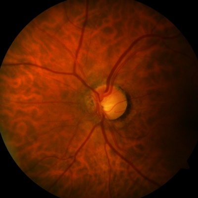
Tilted disc syndrome (TDS), also known as Fuch’s Coloboma, is a congenital anomaly that occurs in 1 to 2% of the population.

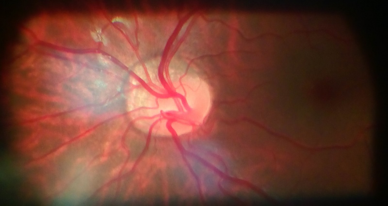
While mostly understood as a nonhereditary process, reports of autosomal dominant inheritance exist. It is characterized by inferonasal tilting of the optic disc and most commonly occurs bilaterally.
Tilted disc syndrome also has an association with high myopia as one-fifth of patients with greater than 5 diopters of myopia have tilted discs.
Bitemporal superior visual field defects are present in about one-fifth of patients with tilted discs and other features of TDS include inferior or inferonasal crescent, the irregular orientation of retinal vessels (situs inversus), and ectasia of the lower fundus or inferior staphyloma.
Tilted disc syndrome is thought to be caused by oblique insertion of the optic nerve and retinal vessels due to incomplete closure of the embryonic fissure of the eye.
Additionally, there is hypoplasia and thinning of the retinal, choroidal, and scleral layers with focal hypopigmentation and ectasia of the inferonasal posterior wall of the globe.
It is unclear whether both the disc anomalies and inferior staphylomas co-exist at birth or whether the inferior staphyloma deepens with time.
Complications
There are various macular complications described in TDS in the literature. These include retinal pigment epithelium (RPE) atrophy, choroidal neovascular membrane (CNVM), subretinal fluid (SRF), polypoidal choroidal vasculopathy (PCV), fovea plana, foveoschisis, and lamellar macular hole (LMH).
Choroidal mechanical and hemodynamic changes at the upper edge of the staphyloma have been proposed to explain the pathophysiology of these complications.
Would you have interest in taking retina images by smartphone?
Fundus photography is superior to fundus analysis as it enables intraocular pathologies to be photo-captured and encrypted information to be shared with colleagues and patients.
Recent technologies allow smartphone-based attachments and integrated lens adaptors to transform the smartphone into a portable fundus camera and Retinal imaging by smartphone.
RETINAL IMAGING BY YOUR SMARTPHONE
REFERENCES
1. Willams, A, et al. The tilted disc syndrome. Practical Neurology, 2005, 5, 54-55
2. Bottoni FG, et al. Dominant inherited tilted disc syndrome and lacquer cracks. Eye (1990), 4, 504-9
3. Manfre L, Vero S, Focarelli-Barone C, Lagall R. Bitemporal Pseudohemianopia related to the “Tilted Disk” Syndrome: CT, MR, and Fundoscopic Findings. AJNR Am J Neuroradiol 20:1750-1751, October 1999
4. Vuori ML Mantyjarvi M. Tilted disc syndrome may mimic false visual field deterioration. Acta Ophthalmol. 2008 Sep;86(6):622-5.
5. Rucker CW (1944) Bitemporal defects in the visual fields resulting from developmental anomalies of the optic discs. Archives of Ophthalmology, 32, 56-9.
RETINAL IMAGING BY YOUR SMARTPHONE

RETINAL IMAGING BY YOUR SMARTPHONE