Papilledema vs papillitis is a common bedside question because both conditions can produce a swollen optic disc.
Papilledema means disc swelling from raised intracranial pressure, while papillitis means anterior optic neuritis.
The disc alone often cannot separate them, so optic nerve function guides the first decision.
Papilledema vs Papillitis at a Glance
- Vision: usually preserved early in papilledema, often reduced acutely in papillitis
- Pain: headache in papilledema, pain on eye movement in papillitis
- Laterality: usually bilateral in papilledema, usually unilateral in adult papillitis
- Pupil: no early RAPD in papilledema, RAPD typical in unilateral papillitis
Vision and Pain in Papilledema vs Papillitis
Central acuity usually stays normal in early papilledema, even when the swelling looks dramatic.
However, patients may report transient visual obscurations lasting seconds.
By contrast, papillitis typically reduces vision over hours to days.
Papillitis also causes an ache that worsens with eye movement, while papilledema tends to cause headache or nausea.
Fundus Explorer Pro
Photograph the retinal findings described here with the phone already in your pocket — 22 D optics and built-in illumination in one handheld unit.
From Choroida — the team behind this sitePupil Findings in Papilledema vs Papillitis
First, check for a relative afferent pupillary defect (RAPD) before dilating the pupils.
Early papilledema usually affects both nerves and spares axonal function, so an RAPD is typically absent.
Unilateral papillitis, however, typically produces a clear RAPD.
Color Vision and Visual Fields
Color desaturation appears early in papillitis, and a red comparison offers a quick bedside test.
Fields often show a central defect.
In papilledema, color vision usually stays normal until late, and perimetry shows an enlarged blind spot.
Disc Appearance
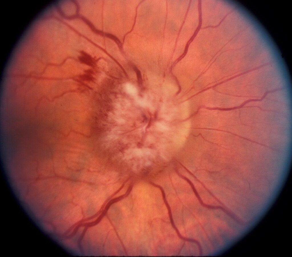
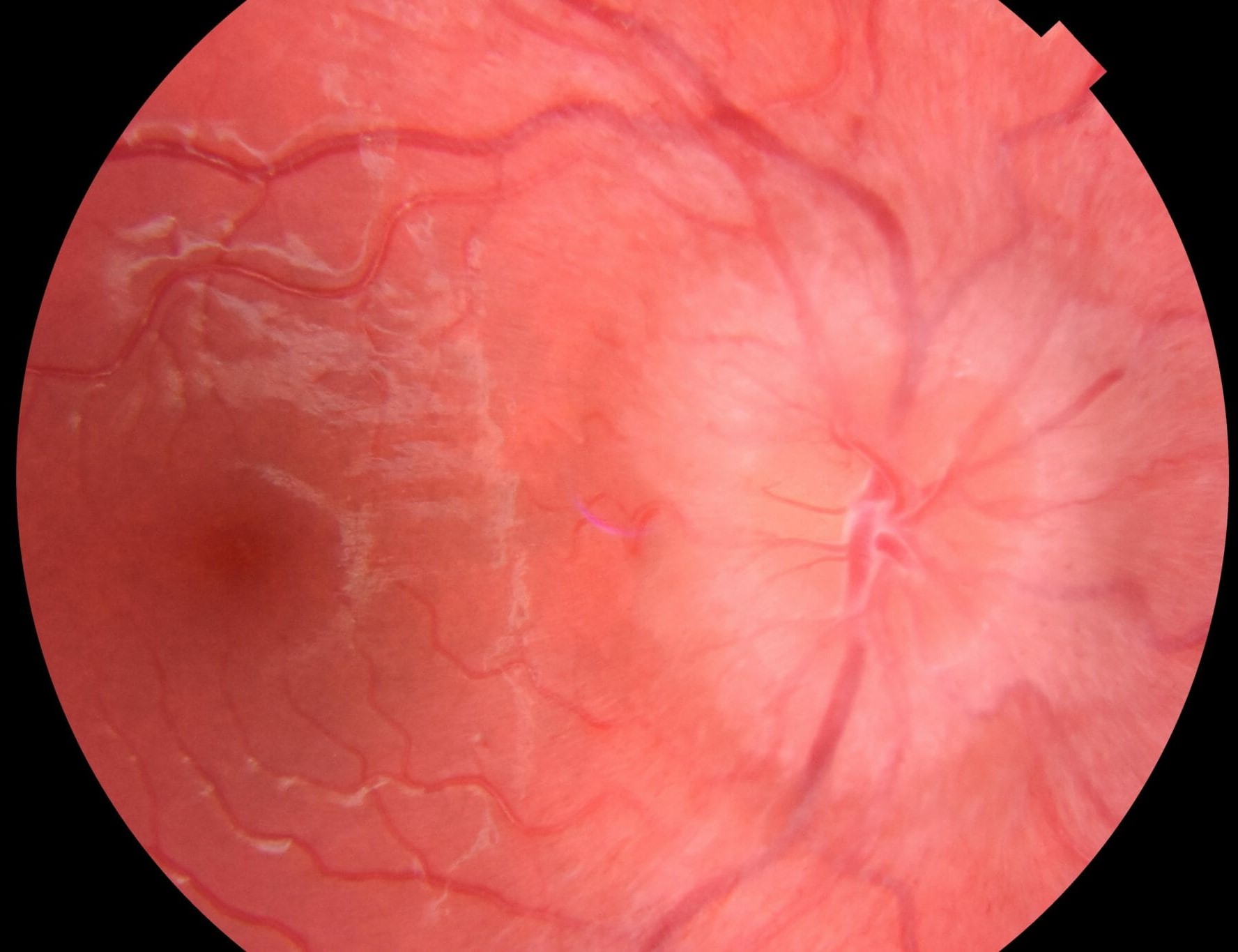
Papilledema often shows circumferential swelling, venous engorgement and hemorrhages as severity increases.
Papillitis usually looks more modest, with diffuse hyperemic swelling.
However, appearance overlaps considerably, so never decide from the disc alone.
Why the Disc Can Look Normal
Most optic neuritis is retrobulbar, and the Optic Neuritis Treatment Trial found disc swelling in about one third of patients.
Therefore, a normal disc does not exclude optic neuritis in a patient with pain and an RAPD.
OCT in Papilledema vs Papillitis
OCT shows peripapillary nerve fiber layer thickening in both conditions, so thickness alone cannot separate them.
It does help document severity and track change between visits.
Examination Pearls for the Swollen Disc
First, look for spontaneous venous pulsations, because their presence makes high intracranial pressure less likely.
However, about one in five normal eyes lacks them, so absence proves nothing.
Also, grade the swelling with the Frisén scale, because a consistent grade makes follow-up comparisons easier.
Finally, examine the fellow eye carefully, because subtle swelling there changes the diagnosis.
Workup for Suspected Papilledema
Treat bilateral disc swelling as urgent and measure blood pressure immediately.
Next, arrange prompt neuroimaging with venography to look for a mass or venous sinus thrombosis.
Lumbar puncture with opening pressure follows imaging when no contraindication exists.
Workup for Papillitis
Contrast MRI of the orbits and brain can show optic nerve enhancement and white matter lesions.
These lesions matter because they strongly influence the later risk of multiple sclerosis.
In addition, atypical cases deserve aquaporin-4 and MOG antibody testing.
Red Flags for Atypical Papillitis
- No pain, or severe pain lasting beyond two weeks
- Bilateral simultaneous loss in an adult
- No recovery after several weeks
- Marked hemorrhages, exudates or a macular star
Treatment in Papilledema vs Papillitis
Papilledema treatment targets the cause of raised pressure.
For idiopathic intracranial hypertension, weight loss and acetazolamide form the usual first line.
The IIHTT showed a modest field benefit in mild visual loss, but paresthesia and fatigue often limit tolerance.
For typical papillitis, the ONTT showed that intravenous methylprednisolone speeds recovery without clearly improving long-term vision.
Oral prednisone alone at standard doses was also linked to more recurrences.
Counseling and Follow-Up
Tell patients with papilledema that good acuity does not guarantee safety, because fields can deteriorate first.
Also, ask them to report worsening obscurations immediately.
Patients with typical papillitis can usually expect good recovery, although subtle color complaints may persist.
In both groups, repeat acuity, fields and disc imaging, because failure to improve should reopen the diagnosis.
Mimics Beyond Papilledema vs Papillitis
Not every elevated disc fits either label (see causes of optic disc swelling).
For example, buried drusen can create pseudopapilledema with no true edema.
A later macular star, meanwhile, should suggest neuroretinitis.
Pitfalls to Avoid
- Do not call bilateral swelling papilledema without measuring blood pressure
- Never delay neuroimaging because acuity is normal
- Do not perform lumbar puncture before imaging
- Avoid labeling painless, non-recovering loss as typical optic neuritis


Document what you see
Two smartphone imaging tools built for everyday clinic use — one for the slit lamp, one for the fundus.
From Choroida — the team behind this siteReferences
- Beck RW, Cleary PA, Anderson MM Jr, et al. A randomized, controlled trial of corticosteroids in the treatment of acute optic neuritis. N Engl J Med. 1992;326:581-588.
- Optic Neuritis Study Group. Multiple sclerosis risk after optic neuritis: final optic neuritis treatment trial follow-up. Arch Neurol. 2008;65:727-732.
- Wall M, McDermott MP, Kieburtz KD, et al. Effect of acetazolamide on visual function in patients with idiopathic intracranial hypertension and mild visual loss. JAMA. 2014;311:1641-1651.
- Friedman DI, Liu GT, Digre KB. Revised diagnostic criteria for the pseudotumor cerebri syndrome in adults and children. Neurology. 2013;81:1159-1165.
- Frisén L. Swelling of the optic nerve head: a staging scheme. J Neurol Neurosurg Psychiatry. 1982;45:13-18.

Writes and reviews the clinical articles on Educate. Every post is checked against peer-reviewed sources before it goes live.