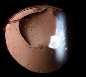
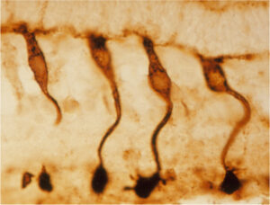
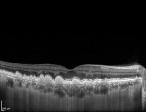
Cloudy cornea describes any loss of corneal clarity from edema, scarring, infiltrate, deposits or abnormal development.
The likely cause changes with the patient’s age.
Therefore, age at onset and one targeted exam clue can narrow the differential quickly.
Reading a Cloudy Cornea at the Slit Lamp
First, identify the affected layer and decide whether the haze represents edema or a solid opacity (see slit-lamp technique).
Edema usually looks diffuse and may come with epithelial bullae or Descemet folds.
In contrast, scars and deposits have sharper borders and do not change during the day.
Next, check laterality, corneal diameter, thickness, sensation, fluorescein staining and intraocular pressure.
Cloudy Cornea in the Newborn
A newborn with corneal haze needs urgent assessment because visual deprivation can cause dense amblyopia.
The mnemonic STUMPED recalls the main causes:
- Sclerocornea
- Tears in Descemet membrane
- Ulcer
- Metabolic disease
- Peters anomaly
- Endothelial dystrophy
- Dermoid
All-fit Slit-Lamp Adapter
Record and share exactly what you see at the slit lamp. One adapter fits any slit lamp or surgical microscope — and any smartphone.
From Choroida — the team behind this sitePrimary Congenital Glaucoma
Enlarged corneas, tearing, photophobia and blepharospasm suggest this diagnosis.
Raised pressure stretches the infant eye and ruptures Descemet membrane.
As a result, Haab striae appear as horizontal or curved lines.
Birth Trauma
Forceps injury typically produces unilateral haze with a vertical or oblique break in Descemet membrane.
However, corneal diameter and pressure usually remain normal.
Although the edema often clears, astigmatism and amblyopia may persist, so follow-up continues.
Peters Anomaly and Sclerocornea
A central opacity with a posterior corneal defect suggests Peters anomaly.
Sclerocornea, however, shows white vascularized tissue that blurs the limbus.
A limbal dermoid forms a raised mass, often inferotemporally.
Congenital Hereditary Endothelial Dystrophy
This condition causes bilateral ground-glass haze with a markedly thick cornea.
However, the thick cornea can falsely elevate pressure readings and mimic glaucoma.
Therefore, interpret tonometry alongside pachymetry and corneal diameter.
Metabolic Cloudy Cornea in Infants
Mucopolysaccharidoses cause progressive stromal clouding that may develop after birth.
Hurler syndrome often clouds the cornea, while Hunter syndrome usually leaves it clinically clear.
Coarse facial features or developmental delay then point toward systemic testing.
Because retinal and optic nerve disease can coexist, a clear graft may not restore good vision.
Cloudy Cornea in Children
Older children more often have acquired disease, so ask about trauma, contact lenses, infection and systemic illness.
Vitamin A deficiency causes xerosis, then keratomalacia in severe cases.
Vernal disease can produce a shield ulcer beneath giant upper tarsal papillae.
Fine refractile crystals with photophobia suggest cystinosis, which also requires renal evaluation.
Herpes simplex may also leave recurrent stromal scars.
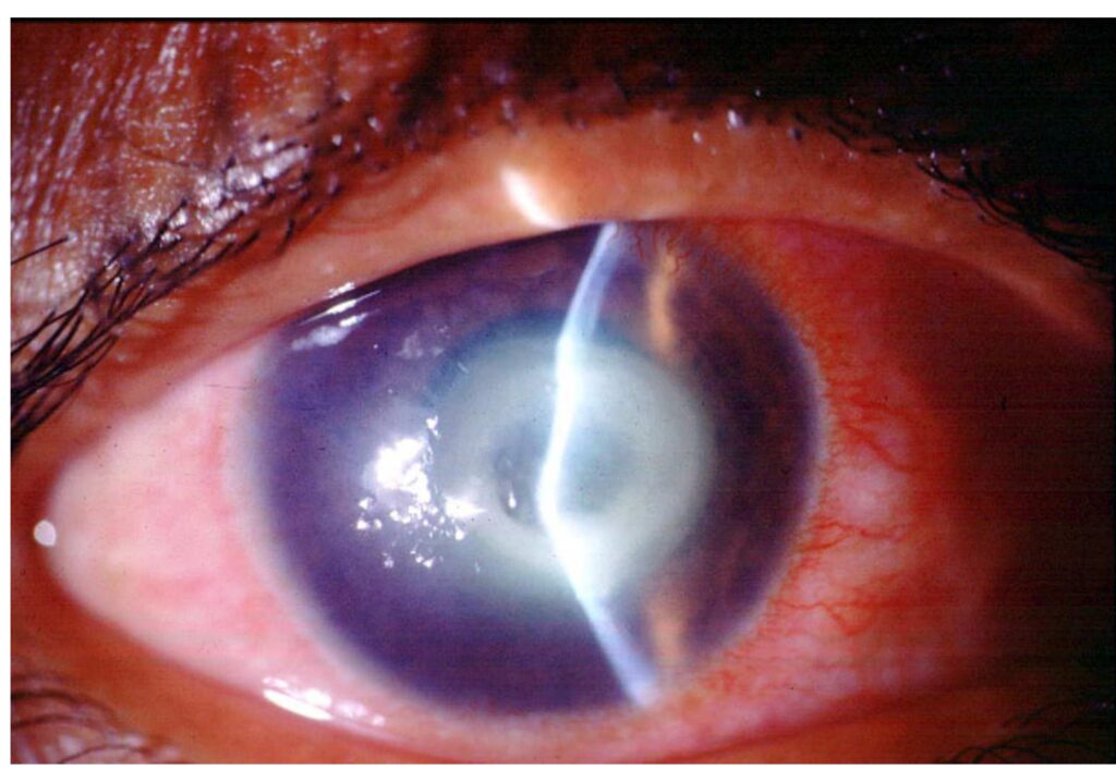
Corneal Edema in Adults
Adults commonly develop haze from endothelial failure.
Fuchs dystrophy shows guttae, and patients describe blur that is worst on waking.
Meanwhile, pseudophakic edema follows surgery.
Sudden central edema in keratoconus suggests hydrops.
The Painful Cloudy Cornea in Adults
Pain changes the priority because infection or high pressure can threaten vision quickly.
A focal white infiltrate with an epithelial defect suggests microbial keratitis until proven otherwise.
On the other hand, a mid-dilated pupil, shallow chamber and very high pressure indicate acute angle closure.
Dystrophies and Deposits
Painless, bilateral opacities with a family history suggest a stromal dystrophy.
Band keratopathy instead forms calcium deposits across the interpalpebral zone, often in chronic uveitis.
Finally, arcus senilis leaves a clear zone at the limbus and rarely affects vision.
Investigations for a Cloudy Cornea
Infants often need examination under anesthesia to measure pressure, corneal diameter and axial length.
When opacity hides the anterior segment, ultrasound biomicroscopy or anterior segment OCT can show iris and lens adhesions.
Adults may also need specular microscopy, pachymetry or cultures.
Treating a Cloudy Cornea
Treatment targets the cause, so congenital glaucoma needs pressure control through angle surgery before graft decisions.
Cysteamine drops can dissolve cystine crystals, while microbial keratitis requires prompt antimicrobial therapy.
Hypertonic saline may reduce morning edema in adults, but it cannot restore endothelial function.
Endothelial keratoplasty then becomes the usual option for visually significant endothelial failure, although rejection and graft failure remain risks.
Counseling Families About Cloudy Cornea
Infant keratoplasty carries a higher failure risk than adult surgery and demands intensive follow-up.
Even with a clear graft, amblyopia, glaucoma or retinal disease may limit vision.
Therefore, parents should expect repeated examinations, refraction and occlusion therapy.
Adults also need clear advice about graft rejection symptoms and when to return urgently.
Pitfalls to Avoid
- Never attribute newborn corneal haze to birth trauma without measuring pressure
- Do not rely on tonometry alone when the cornea is abnormally thick
- Avoid labeling an infiltrate as a scar before checking fluorescein staining
- Do not delay referral when the opacity covers the visual axis in an infant


Document what you see
Two smartphone imaging tools built for everyday clinic use — one for the slit lamp, one for the fundus.
From Choroida — the team behind this siteReferences
- Nischal KK. Congenital corneal opacities: a surgical approach to nomenclature and classification. Eye (Lond). 2007;21:1326-1337.
- Weiss JS, Moller HU, Aldave AJ, et al. IC3D classification of corneal dystrophies, edition 2. Cornea. 2015;34:117-159.
- Ashworth JL, Biswas S, Wraith E, Lloyd IC. Mucopolysaccharidoses and the eye. Surv Ophthalmol. 2006;51:1-17.
- Whitcher JP, Srinivasan M, Upadhyay MP. Corneal blindness: a global perspective. Bull World Health Organ. 2001;79:214-221.
- American Academy of Ophthalmology. Basic and Clinical Science Course, Section 8: External Disease and Cornea. San Francisco: AAO.
- Bowling B. Kanski’s Clinical Ophthalmology. 8th ed. Elsevier; 2016.

Writes and reviews the clinical articles on Educate. Every post is checked against peer-reviewed sources before it goes live.