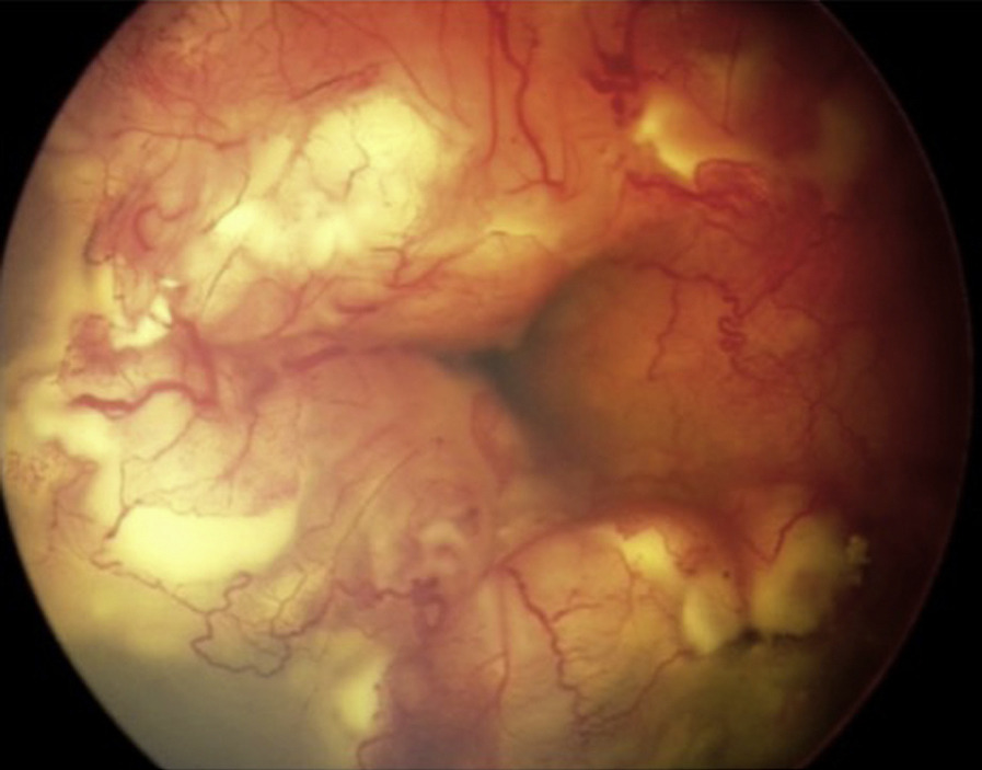
Retinoblastoma prognosis covers three separate outcomes: survival, eye salvage and useful vision.
In high-income countries, nearly every child now survives the disease. However, survival falls sharply where diagnosis comes late and treatment is hard to reach, and families need each outcome explained on its own, because the odds differ.
Survival in Retinoblastoma Prognosis
Retinoblastoma is highly curable while it stays inside the globe, and the 2020 global study found near-universal survival in high-income countries. In contrast, survival was much lower in low-income countries, where late, often extraocular, disease at presentation drove most of that gap.
Stage at Diagnosis
Stage at diagnosis is the strongest single predictor of outcome. A child with leukocoria and a small intraocular tumor has an excellent outlook (see retinoblastoma), while proptosis, orbital extension or metastasis change the picture completely. As a result, a delayed referral can cost a life as well as an eye.
Fundus Explorer Pro
Photograph the retinal findings described here with the phone already in your pocket — 22 D optics and built-in illumination in one handheld unit.
From Choroida — the team behind this siteClassification Systems and Retinoblastoma Prognosis
The international intraocular classification groups eyes from A to E, and it mainly predicts the chance of saving the eye. The AJCC eighth edition also adds TNM staging and a heritable trait category, so use both systems when you counsel parents.
Eye Salvage by Group
Groups A to C usually respond well to systemic chemoreduction and focal therapy, while group D eyes have lower salvage rates with intravenous chemotherapy alone. However, intra-arterial chemotherapy has improved salvage in many advanced eyes. Group E eyes usually need enucleation, because hidden high-risk features are common.
Vision Outcomes
Final acuity depends mostly on how close the tumor sits to the fovea and disc. A small peripheral tumor treated early may leave near-normal vision, but macular tumors leave scars that limit central acuity even after cure. Amblyopia, cataract and retinal detachment can reduce vision further, so patch the better eye when the treated eye still has potential.
High-Risk Pathology in Retinoblastoma Prognosis
After enucleation, histopathology defines the metastatic risk. Recognized high-risk features include:
- Massive choroidal invasion (3 mm or more)
- Postlaminar optic nerve invasion
- Scleral or extrascleral extension
- Anterior segment invasion
- Tumor at the optic nerve resection margin
Adjuvant Therapy
Children with high-risk features usually receive adjuvant chemotherapy, and Honavar and colleagues found fewer metastases among treated children. A positive resection margin often prompts orbital radiotherapy as well. Therefore, wait for full pathology before giving a final prognosis.
Extraocular and Metastatic Disease
Orbital disease needs intensive multimodal treatment, and some children are cured. However, central nervous system spread carries a very poor outlook. Distant metastasis may respond to high-dose chemotherapy with stem cell rescue, although results vary between centers.
Genetics and Retinoblastoma Prognosis
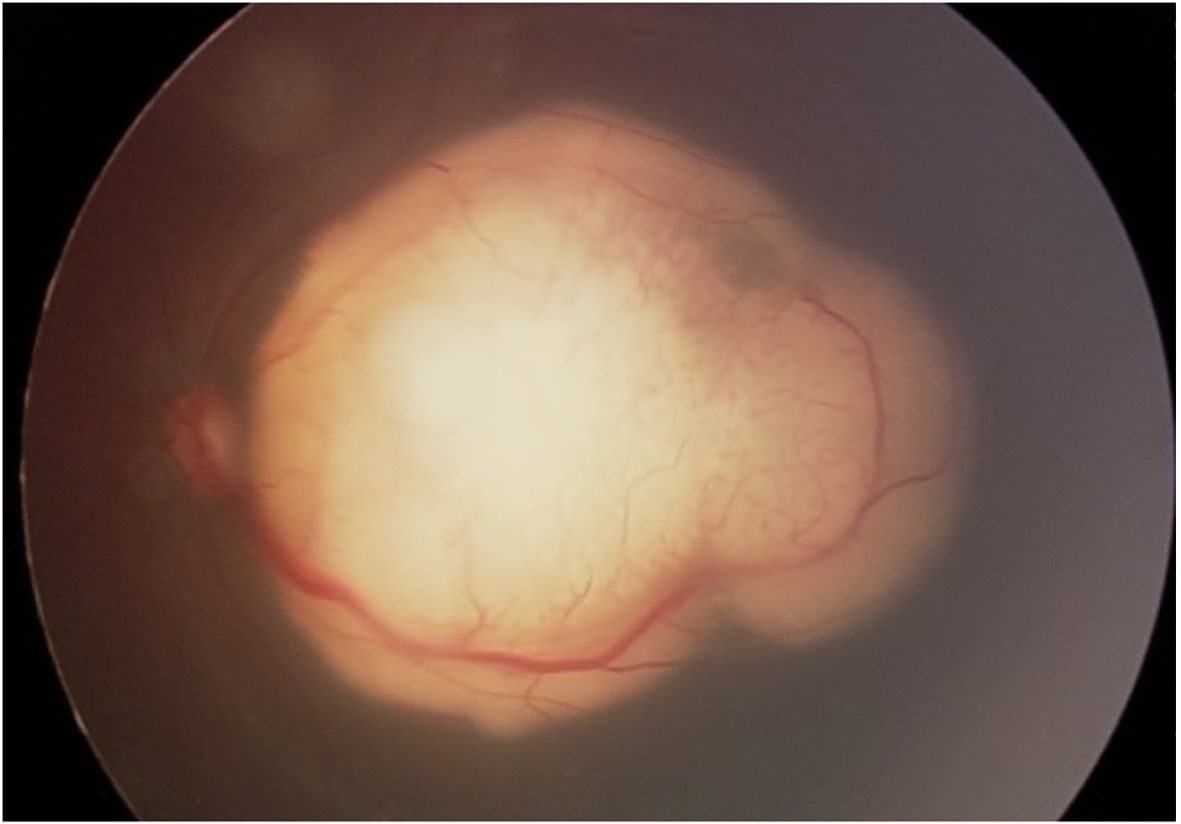
Heritable disease carries a germline RB1 variant, and these children often have bilateral or multifocal tumors, while new tumors may appear in either eye during early childhood. A few develop a midline intracranial tumor, which carries a guarded outlook.
Second Cancers in Survivors
Survivors with heritable disease face a lifelong risk of second cancers, and sarcomas of bone and soft tissue, along with melanoma, are well described. External beam radiation raises the risk, particularly within the treated field. As a result, modern protocols avoid radiation whenever possible.
Unilateral Versus Bilateral Disease
Most unilateral cases are nonheritable, yet some carry a germline variant, so blood and tumor testing should clarify the risk. Bilateral cases often present younger, especially when family history prompts screening, and screening siblings and offspring finds tumors at a treatable stage.
Treatment Complications
Every therapy carries a cost, and intra-arterial chemotherapy may cause choroidal or retinal vascular occlusion. Systemic carboplatin can cause marrow suppression and hearing loss. Intravitreal melphalan helps control vitreous seeds, while it may cause retinal toxicity.
Retinoblastoma Prognosis and Access to Care
Global outcomes track national income closely, and families in low-resource settings often present late and may abandon treatment. For example, leukocoria awareness campaigns can shift the stage at diagnosis.
Counseling Families on Retinoblastoma Prognosis
Start with the outcome the family fears most, usually survival, then explain eye salvage and vision as separate questions. Quote ranges from your own center when available, and avoid single percentages from distant series, because they rarely fit the individual child. Finally, revisit the discussion when pathology returns.
Follow-Up and Retinoblastoma Prognosis
Treated eyes need examinations under anesthesia until regression looks stable, and recurrence is most likely in the early period after therapy. Later, survivors need long-term review of vision, hearing and second cancers.
Pitfalls to Avoid
- Do not quote eye salvage figures as survival figures
- Never promise vision before assessing the macula
- Do not skip genetic testing in unilateral cases
- Avoid radiation in heritable disease when safer options exist
- Do not relax surveillance once the first eye is stable


Document what you see
Two smartphone imaging tools built for everyday clinic use — one for the slit lamp, one for the fundus.
From Choroida — the team behind this siteReferences
- Global Retinoblastoma Study Group, Fabian ID, Abdallah E, et al. Global retinoblastoma presentation and analysis by national income level. JAMA Oncol. 2020;6:685-695.
- Kaliki S, Shields CL, Rojanaporn D, et al. High-risk retinoblastoma based on International Classification of Retinoblastoma: analysis of 519 enucleated eyes. Ophthalmology. 2013;120:997-1003.
- Honavar SG, Singh AD, Shields CL, et al. Postenucleation adjuvant therapy in high-risk retinoblastoma. Arch Ophthalmol. 2002;120:923-931.
- Kleinerman RA, Tucker MA, Tarone RE, et al. Risk of new cancers after radiotherapy in long-term survivors of retinoblastoma: an extended follow-up. J Clin Oncol. 2005;23:2272-2279.
- Abramson DH, Dunkel IJ, Brodie SE, et al. A phase I/II study of direct intraarterial (ophthalmic artery) chemotherapy with melphalan for intraocular retinoblastoma: initial results. Ophthalmology. 2008;115:1398-1404.
- Dimaras H, Corson TW, Cobrinik D, et al. Retinoblastoma. Nat Rev Dis Primers. 2015;1:15021.

Writes and reviews the clinical articles on Educate. Every post is checked against peer-reviewed sources before it goes live.