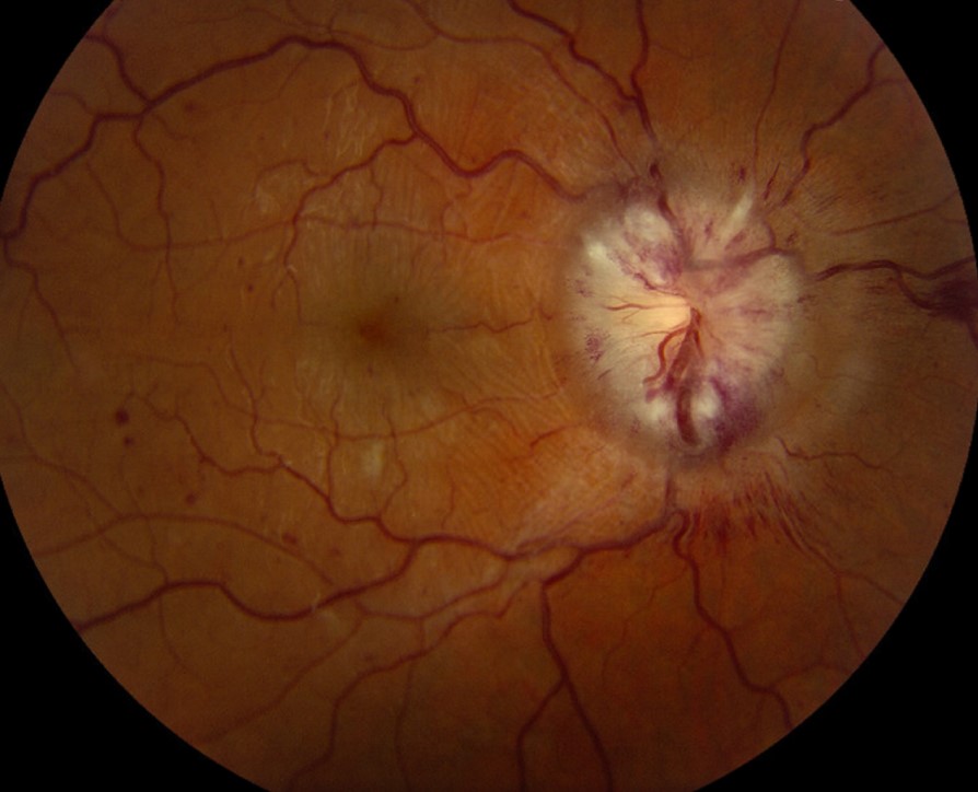
Dragged disc describes an optic disc and retinal vessels that appear pulled toward a contracting retinal scar.
Clinicians usually meet it as a late sign of retinopathy of prematurity (ROP) or familial exudative vitreoretinopathy (FEVR).
Because the same traction can displace the fovea, the sign also prompts a careful macular assessment.
What a Dragged Disc Looks Like
The disc looks pulled toward one side, and its vessels leave at an unusual angle.
In the common temporal pattern, the arcades lose their curve and run straighter across the macula.
In addition, a retinal fold sometimes leads from the disc toward the peripheral scar.
Why a Dragged Disc Develops
Peripheral fibrovascular tissue contracts as active disease regresses.
As a result, the scar pulls the retina and its vessels toward itself.
The nerve head generally stays in position, so “dragged” mainly describes the overlying retina and vessels.
Fundus Explorer Pro
Photograph the retinal findings described here with the phone already in your pocket — 22 D optics and built-in illumination in one handheld unit.
From Choroida — the team behind this siteDragged Disc After ROP
Regressed ROP is the classic cause in a former preterm infant.
The temporal retina usually hosts the ridge, so traction tends to pull vessels temporally.
Therefore, ask about gestational age, birth weight, neonatal oxygen and earlier treatment.
Dragged Disc in FEVR
FEVR produces a similar picture, often in a patient born at full term.
Incomplete peripheral vascularization drives exudation, neovascularization and traction.
Relatives may also carry it without symptoms (see FEVR overview).
Other Causes of a Dragged Disc
Other causes include:
- Ocular toxocariasis, especially with a peripheral granuloma and fibrous band
- Persistent fetal vasculature with a stalk or retinal fold
- Incontinentia pigmenti with peripheral nonperfusion and scarring
- Combined hamartoma of the retina and retinal pigment epithelium near the disc
- Scarring after trauma, uveitis or sickle retinopathy
What Patients Report
Many patients have no symptoms, and a routine exam finds the sign by chance.
Parents may instead notice an eye turn or poor fixation.
Older patients may report floaters or a shadow because a tear or detachment has developed.
Examining a Dragged Disc
First, compare both discs and trace the arcades toward their peripheral endpoints.
Then locate the fovea relative to the disc, looking for macular heterotopia.
Next, examine the periphery for avascular retina, ridge remnants, pigment and retinal breaks.
Finally, check refraction, alignment and anterior chamber depth, since regressed ROP can accompany a shallow chamber.
Imaging the Traction
Widefield color photography documents the vessel course and scar location.
Fluorescein angiography then shows peripheral nonperfusion, leakage and abrupt vessel endings that examination may miss.
OCT adds macular detail, including traction, folds and a shallow foveal pit.
Effect on Vision
Vision ranges from normal to severely reduced, so the sign alone cannot predict acuity.
Foveal structure usually tells you more than disc appearance.
Macular folds and persistent traction can limit acuity, while myopia, anisometropia and amblyopia may add a treatable component in children.
Apparent Exotropia
A temporally displaced fovea makes the eye rotate outward to fixate.
As a result, the eye can look exotropic, although cover testing shows no refixation movement.
However, true strabismus also occurs in these patients, so always perform the cover test.
Differential Diagnosis of Dragged Disc
A tilted disc shows an oblique axis and often an inferonasal crescent, without peripheral traction.
An optic disc coloboma shows inferior excavation, which a dragged disc lacks.
Blurred margins can also suggest disc edema, although straightened vessels and a peripheral scar favor traction.
Managing a Dragged Disc
No treatment reliably returns the vessels to their original position.
Therefore, management targets the underlying disease and its complications.
Active ROP follows established criteria, including those from the ETROP trial.
In FEVR, clinicians often laser avascular retina when leakage or neovascularization appears, although controlled evidence is limited.
When Surgery Helps
Vitrectomy or scleral buckling may relieve progressive traction or repair a detachment.
However, surgery aims to stabilize the retina, and it rarely reverses long-standing heterotopia.
Risks include retinal breaks, cataract and redetachment.
Stable, asymptomatic dragging therefore usually needs observation alone.
Counseling Patients
Explain that the appearance reflects an old scar and that vision depends mainly on the fovea.
Also teach the warning symptoms of detachment: new floaters, flashes or a shadow.
Children need glasses and amblyopia treatment when indicated, while relatives of FEVR patients may need examination and genetic counseling.
Follow-Up for Dragged Disc
Long-term follow-up suits most patients because tears and detachments can occur years later.
Repeat angiography when you suspect new leakage or neovascularization.
Pitfalls to Avoid
- Prematurity does not explain every case, because FEVR can look identical
- Check angle kappa and cover testing before planning strabismus surgery
- Never stop at the disc, because the cause usually lies in the periphery
- Examine family members when FEVR is plausible


Document what you see
Two smartphone imaging tools built for everyday clinic use — one for the slit lamp, one for the fundus.
From Choroida — the team behind this siteReferences
- Chiang MF, Quinn GE, Fielder AR, et al. International Classification of Retinopathy of Prematurity, Third Edition. Ophthalmology. 2021;128:e51-e68.
- Early Treatment for Retinopathy of Prematurity Cooperative Group. Revised indications for the treatment of retinopathy of prematurity. Arch Ophthalmol. 2003;121:1684-1694.
- Criswick VG, Schepens CL. Familial exudative vitreoretinopathy. Am J Ophthalmol. 1969;68:578-594.
- Ranchod TM, Ho LY, Drenser KA, Capone A, Trese MT. Clinical presentation of familial exudative vitreoretinopathy. Ophthalmology. 2011;118:2070-2075.
- Goldberg MF. Persistent fetal vasculature (PFV): an integrated interpretation of signs and symptoms. Am J Ophthalmol. 1997;124:587-626.

Writes and reviews the clinical articles on Educate. Every post is checked against peer-reviewed sources before it goes live.