Persistent subretinal fluid is fluid that remains under the macula after an anatomically successful retinal detachment repair.
The break is closed and the retina looks attached, yet OCT still shows a shallow detachment. As a result, patients often notice that vision lags behind their expectations. Clinicians should therefore recognize the pattern and explain it early.
What Persistent Subretinal Fluid Means
The term describes fluid seen on OCT weeks after surgery with no open retinal break. It is often subtle on biomicroscopy, so many cases surfaced only once OCT became routine, and it also differs from a recurrent detachment, which needs further surgery.
Which Surgeries Leave Fluid Behind
Persistent fluid appears most often after scleral buckling, especially without external drainage, and it also follows pneumatic retinopexy, because both procedures rely on the eye to absorb residual fluid. After vitrectomy, internal drainage and gas tamponade usually clear the fluid, although small blebs can still appear (see scleral buckle vs vitrectomy).
Fundus Explorer Pro
Photograph the retinal findings described here with the phone already in your pocket — 22 D optics and built-in illumination in one handheld unit.
From Choroida — the team behind this siteRisk Factors for Persistent Subretinal Fluid
Several patient and detachment features raise the risk.
- Younger age with formed, attached vitreous
- Macula-off detachment of long duration
- Chronic detachment with demarcation lines or subretinal precipitates
- Scleral buckle without external drainage
- Pneumatic retinopexy
- Shallow, slowly progressive inferior detachment
Why the Fluid Persists
The exact mechanism is still debated, but fluid in chronic detachments is often viscous and rich in protein, so the retinal pigment epithelium pumps it out slowly. In addition, the RPE may stay dysfunctional after long separation from the photoreceptors, and debris from shed outer segments may also thicken the fluid.
OCT Appearance of Persistent Subretinal Fluid
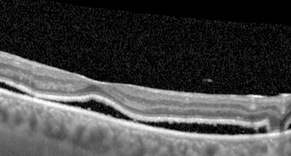
OCT shows a dome-shaped hyporeflective space between the neurosensory retina and the RPE, which may form one subfoveal pocket or several separate blebs around the fovea. The overlying outer retina often looks thickened, with hyperreflective dots at the photoreceptor tips (for a broader guide, see subretinal fluid on OCT).
Blebs or a Single Pocket
Some eyes show a single shallow pocket under the fovea, while others show several small blebs, often called multiple subretinal fluid blebs. Then each bleb tends to shrink at its own pace.
Exam Findings in Persistent Subretinal Fluid
Clinical examination is often unremarkable, although a careful look may show a subtle yellowish elevation or a blunted foveal reflex. Amsler grid testing may also reveal distortion, and OCT therefore confirms the diagnosis.
Symptoms and Visual Effects
Patients report blur, metamorphopsia, micropsia or a dim central patch, and visual acuity often improves more slowly than expected. Because the photoreceptors stay separated from the RPE, their recovery is delayed, and OCT studies have linked subfoveal fluid with incomplete early recovery after macula-off detachments.
Expected Resolution Time for Persistent Subretinal Fluid
Most persistent fluid resolves spontaneously, but the timeline varies widely, from weeks to many months. Some reports describe fluid lasting beyond a year, mainly in young patients after buckling, so serial OCT is the best way to track its course.
Final Visual Outcome
Once the fluid absorbs, vision usually continues to improve, and in many series long-term acuity ends up similar to eyes without persistent fluid. However, other studies suggest a modest effect on final vision, so the evidence is mixed. Outer retinal integrity on OCT, especially the ellipsoid zone, also helps predict recovery.
What Must Be Excluded
Before calling the fluid benign, exclude conditions that need action.
- Recurrent detachment from a new or reopened break (see recurrent retinal detachment after vitrectomy)
- An open break on the buckle
- Cystoid macular edema
- Epiretinal membrane with traction
- Choroidal effusion after buckling
- Central serous chorioretinopathy in steroid users
Management of Persistent Subretinal Fluid
Observation is the standard approach when the retina is attached and no break is open, and no treatment reliably speeds absorption in controlled studies. Some surgeons have tried topical or oral agents; however, evidence comes mainly from small reports. Therefore, avoid repeat surgery for fluid alone unless the retina detaches again.
Follow-Up Plan
Repeat OCT at regular intervals until the fluid clears, then compare scans for bleb height and extent. Also examine the periphery at each visit to confirm the breaks remain sealed. Any increase in fluid should prompt a full search for a new break.
Counseling Patients With Persistent Subretinal Fluid
Explain that the retina is attached and the surgery has worked. Next, describe the fluid as a slow-draining pocket the eye clears by itself, and set realistic expectations about months of gradual change. Finally, teach warning signs such as new floaters, flashes or a spreading shadow.
Pitfalls to Avoid
- Do not label every fluid pocket as a failed repair
- Never skip the peripheral exam when OCT shows fluid
- Avoid promising a fixed date for clearance
- Do not ignore new distortion between scheduled visits


Document what you see
Two smartphone imaging tools built for everyday clinic use — one for the slit lamp, one for the fundus.
From Choroida — the team behind this siteReferences
- Hagimura N, Iida T, Suto K, Kishi S. Persistent foveal retinal detachment after successful rhegmatogenous retinal detachment surgery. Am J Ophthalmol. 2002;133:516-520.
- Benson SE, Schlottmann PG, Bunce C, Xing W, Charteris DG. Optical coherence tomography analysis of the macula after scleral buckle surgery for retinal detachment. Ophthalmology. 2007;114:108-112.
- Seo JH, Woo SJ, Park KH, Yu YS, Chung H. Influence of persistent submacular fluid on visual outcome after successful scleral buckle surgery for macula-off retinal detachment. Am J Ophthalmol. 2008;145:915-922.
- Wolfensberger TJ, Gonvers M. Optical coherence tomography in the evaluation of incomplete visual acuity recovery after macula-off retinal detachments. Graefes Arch Clin Exp Ophthalmol. 2002;240:85-89.
- American Academy of Ophthalmology. Basic and Clinical Science Course, Section 12: Retina and Vitreous. San Francisco: AAO; 2023.
Test yourself
A few questions straight from this article.
-
How is persistent subretinal fluid after retinal detachment repair defined?
It is fluid seen on OCT weeks after surgery with the break closed and the retina attached, which separates it from a recurrent detachment needing further surgery. -
After which retinal detachment repair is persistent subretinal fluid seen most often?
Buckling without drainage and pneumatic retinopexy rely on the eye to absorb residual fluid, whereas vitrectomy drains it internally. -
Which patient feature raises the risk of persistent subretinal fluid after detachment repair?
Young age with formed vitreous, long-standing macula-off or chronic detachment, and shallow slowly progressive inferior detachments all raise the risk. -
What is the leading explanation for why persistent subretinal fluid absorbs slowly after detachment repair?
Fluid in chronic detachments is often viscous and protein-rich, and an RPE long separated from photoreceptors may stay dysfunctional, so clearance is slow. -
What does OCT typically show in persistent subretinal fluid after detachment repair?
The fluid sits between the neurosensory retina and the RPE as a single subfoveal pocket or several blebs, often with thickened outer retina and hyperreflective dots. -
What does clinical examination most often show in persistent subretinal fluid after detachment repair?
Examination may show only a subtle yellowish elevation or blunted foveal reflex, which is why OCT confirms the diagnosis. -
What is the expected course of persistent subretinal fluid after successful detachment repair?
Most persistent fluid clears on its own, though timing varies widely and some cases last beyond a year, mainly in young patients after buckling. -
Which OCT feature helps predict visual recovery once persistent subretinal fluid absorbs?
Outer retinal integrity on OCT, especially the ellipsoid zone, helps predict recovery, while evidence on long-term acuity effects is mixed. -
What is the standard management of persistent subretinal fluid when the retina is attached and no break is open?
No treatment reliably speeds absorption in controlled studies, so repeat surgery for fluid alone is avoided unless the retina detaches again. -
During follow-up of persistent subretinal fluid, what should an increase in the fluid prompt?
Periphery should be examined at every visit to confirm breaks remain sealed, and any rise in fluid should trigger a search for a new break.

Writes and reviews the clinical articles on Educate. Every post is checked against peer-reviewed sources before it goes live.