
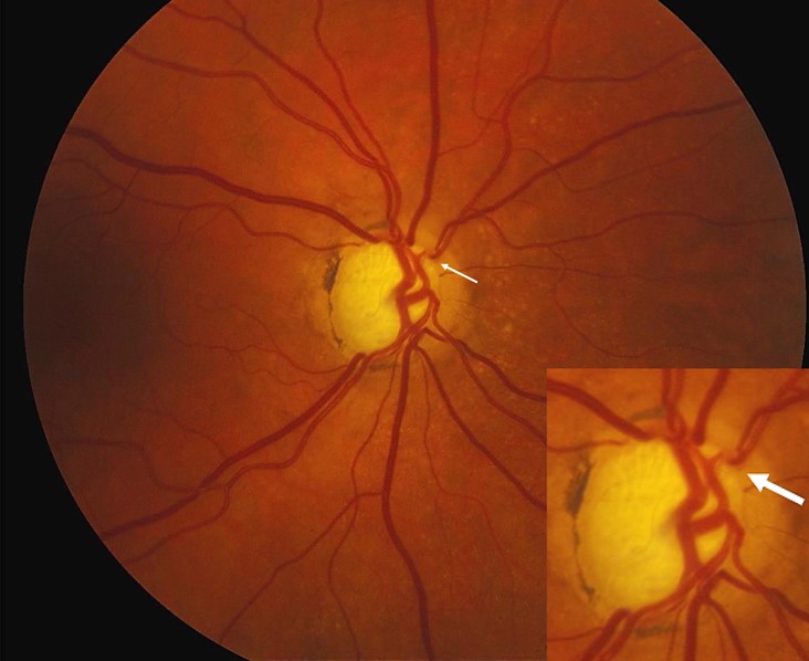
Bayoneting is the sharp bend of a retinal vessel as it crosses the margin of an excavated optic cup, and together with nasalization of the vessel trunk it is a sign of neuroretinal rim loss.
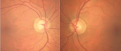
These vascular signs are easy to overlook, because attention is usually drawn to the cup-to-disc ratio.
They provide supporting evidence of glaucomatous optic neuropathy, and they help to monitor change over time.
What Is Bayoneting, and Which Other Vascular Signs Matter?
As the neuroretinal rim thins, the structure of the disc changes, and the vessels that lie on and around it move or bend to follow the new contour.
- Bayoneting: a vessel that passes over the edge of the cup and plunges steeply, with a bend at the rim and another at the base, like a bayonet
- Nasalization: displacement of the central vessel trunk toward the nasal side of the cup
- Baring of circumlinear vessels: a small gap between the vessel and the rim, where the rim has receded and left the vessel bare
- Overpass vessels: a vessel that crosses the excavated cup without touching its base
- Laminar dot sign: visible pores of the lamina cribrosa as the cup deepens
No single sign is specific, and each should be interpreted together with the rim, the nerve fiber layer, and the visual field.
Mechanisms
The vessels are anchored at the lamina and at the retinal surface, and they are rigid compared with the tissue around them.
When the rim tissue is lost and the lamina is displaced backward, the vessels are pulled and bent.
The central retinal vessel trunk moves nasally in many eyes with glaucoma, possibly because the temporal rim has thickened initially and the tissue loss is greater on the other side.
Armaly and later authors have measured vessel displacement as a feature of the normal and the glaucomatous cup, and nasalization is more pronounced in larger cups.
Fundus Explorer Pro
Photograph the retinal findings described here with the phone already in your pocket — 22 D optics and built-in illumination in one handheld unit.
From Choroida — the team behind this siteHow to Examine the Disc
Examine the disc with a dilated pupil and a stereoscopic view, using a 78 or 90 diopter lens at the slit lamp.
Look first at the size of the disc, because large discs have large physiologic cups and small discs may have little cup.
Evaluate the rim for thinning, notching, and pallor.
Follow the vessels from the retinal surface across the rim and into the cup, and note any sharp bend.
Compare with the fellow eye, since glaucoma is often asymmetric.
Record the findings in a drawing and with photographs.
Differential Diagnosis
- Physiologic cupping: a large, symmetric cup with a healthy rim and no vascular distortion
- Optic atrophy of other causes: pallor of the rim with a small cup, and different vessel pattern
- Myopic tilted disc: an oval disc with a temporal crescent and displaced vessels (see tilted disc syndrome)
- Congenital disc anomalies: pits, colobomas, and large discs with different vessel patterns
Using the Signs in Practice
Treat the vascular signs as evidence that the disc has changed, and do not use them alone to decide on treatment.
Support the diagnosis with the intraocular pressure, the angle, the visual field, and OCT of the nerve fiber layer.
A baseline photograph is valuable, and it is the best way to detect a change such as new bayoneting or a wider bare vessel over time.
Patients with a family history, high pressure, or thin corneas who have these signs need careful follow-up (see primary open-angle glaucoma).
Documenting Change Over Time
Vascular signs are valuable because they are easy to compare on serial photographs.
Keep a stereo disc photograph or a good monocular image at baseline, and compare it with images taken every one to two years in patients with glaucoma.
Look for a vessel that was previously touching the rim and now lies away from it, or for a bend that has become more pronounced.
These changes may precede changes in the visual field, and they provide an additional line of evidence that the disease is progressing.
Caveats
In very large discs with deep physiologic cups, some vessel displacement is present from the start, so a single examination can be misleading.
In high myopia, the vessels and the disc are distorted by the shape of the eye, and the signs are harder to interpret.
OCT of the rim and the nerve fiber layer helps in these eyes, because it measures the tissue directly and does not depend on vessel position.
In children and young adults with large cups, take a family history and measure the pressure, and compare the findings with those of the parents when it is practical.


Document what you see
Two smartphone imaging tools built for everyday clinic use — one for the slit lamp, one for the fundus.
From Choroida — the team behind this siteReferences
- Jonas JB, Fernández MC, Stürmer J. Pattern of glaucomatous neuroretinal rim loss. Ophthalmology. 1993;100:63-68.
- Armaly MF. The optic cup in the normal eye. I. Cup width, depth, vessel displacement, ocular tension and outflow facility. Am J Ophthalmol. 1969;68:401-407.
- Shields’ Textbook of Glaucoma. Allingham RR, Damji KF, Freedman S, et al, eds. 6th ed. Lippincott Williams and Wilkins; 2011.
- American Academy of Ophthalmology. Basic and Clinical Science Course, Section 10: Glaucoma. AAO; current edition.

Writes and reviews the clinical articles on Educate. Every post is checked against peer-reviewed sources before it goes live.


