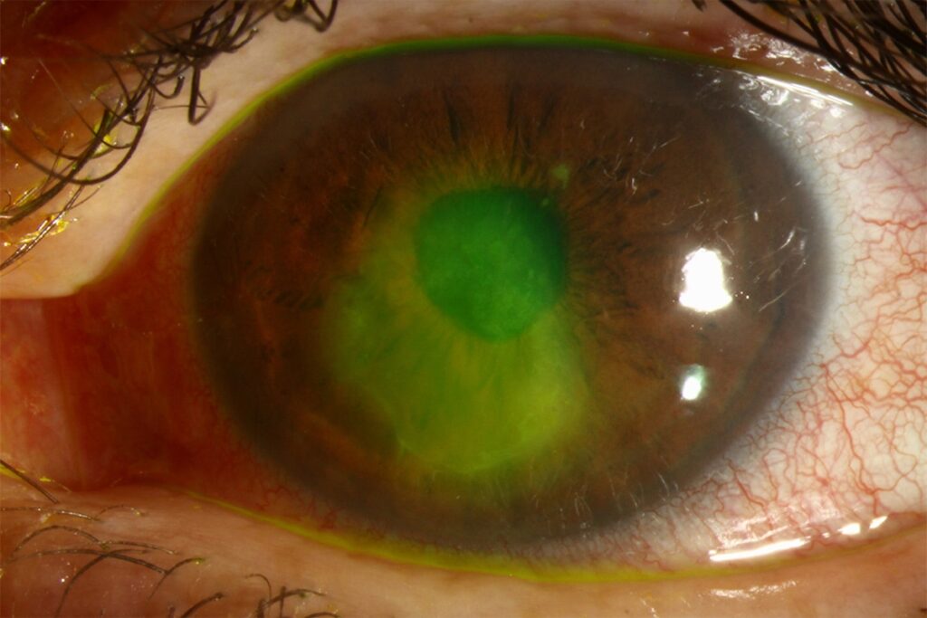
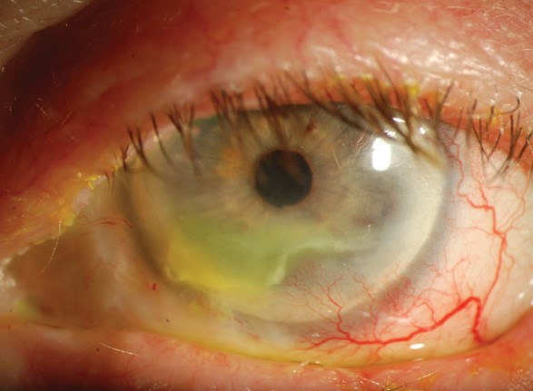
A persistent epithelial defect is a break in the corneal epithelium that does not close within about two weeks of standard treatment.
It can lead to ulceration, infection, melting, and scarring, so the cause must be found and treated.
A stepwise plan, from the simplest measures to surgery, resolves most cases.
Why a Persistent Epithelial Defect Forms
The corneal epithelium normally closes a defect within days, by cell migration from the edge and from stem cells at the limbus.
Also, healing needs healthy nerves, a stable tear film, intact limbal stem cells, and a clean basement membrane.
In addition, loss of any of these can stall the repair (see neurotrophic keratitis).
Persistent Epithelial Defect: Common Causes
- Neurotrophic disease, from herpes, zoster, diabetes, trigeminal damage, or surgery (see corneal sensation testing)
- Dry eye and exposure, with lagophthalmos or poor blinking (see exposure keratopathy)
- Limbal stem cell deficiency, from burns or inflammation (see limbal stem cell deficiency)
- Toxic drops, such as preserved glaucoma drugs and topical anesthetic abuse (see toxic epitheliopathy)
- Autoimmune and inflammatory diseases, such as rheumatoid arthritis and pemphigoid
- Diabetes, with poor healing and abnormal basement membrane
- Recurrent erosions, infection, and surgery such as vitrectomy or corneal procedures
More than one factor, however, is often present.
All-fit Slit-Lamp Adapter
Record and share exactly what you see at the slit lamp. One adapter fits any slit lamp or surgical microscope — and any smartphone.
From Choroida — the team behind this siteEvaluation for Persistent Epithelial Defect
- A careful history of previous disease, surgery, drugs, and systemic conditions
- Corneal sensation testing, and examination of the lids, blink, and closure
- Fluorescein staining, which shows smooth, rolled edges in an old defect
- Cultures when infection is suspected
- Tear tests and lid assessment
The defect often has a gray, heaped edge of epithelium that does not migrate.
Step 1: Remove Insults and Lubricate
- Stop toxic and preserved drops, and then switch to preservative-free formulas
- Use preservative-free lubricants and ointment, often several times a day and at night
- Treat lid disease and exposure, with taping or lid closure at night
- Manage underlying infection or inflammation
Step 2: Protect the Surface in Persistent Epithelial Defect
- Bandage contact lens: protects against lid friction (see bandage contact lens)
- Punctal occlusion: keeps tears on the surface
- Patching or tarsorrhaphy, temporary or permanent, for exposure or severe cases
- Scleral lenses: hold a reservoir of fluid over the cornea (see scleral lens fitting)
Step 3: Biological Therapies
- Autologous serum drops: provide growth factors and nutrients, and are used at concentrations of 20 to 50 percent (see autologous serum eye drops)
- Platelet-rich plasma and cord blood serum: alternatives
- Cenegermin: recombinant human nerve growth factor, shown in randomized trials to heal a significantly larger share of stage 2 and 3 neurotrophic defects by eight weeks than vehicle (see the references)
- Amniotic membrane: as a patch or a ring, which promotes healing and reduces inflammation (see amniotic membrane transplantation)
Step 4: Surgery
- Conjunctival flap: for severe disease, with a good vascular supply, though the vision is lost over the covered area
- Limbal stem cell transplantation for deficiency
- Nerve transfer or corneal neurotization, for selected neurotrophic cases
- Keratoplasty, which is a last resort
- Treatment of perforation, with glue or grafts (see corneal perforation glue)
Monitoring
- See the patient every few days at the start, and then review the size of the defect with a photo
- Look for infection, thinning, and melting
- Avoid steroids unless the cause is inflammatory, and use them with great care
- Continue treatment after the defect closes, to avoid recurrence
Patients should also know the symptoms that need urgent review.
A Typical Case
A 62-year-old man with long-standing diabetes and a previous vitrectomy has a central epithelial defect of four millimeters that has not healed after three weeks of lubricants and a bandage lens.
Corneal sensation is also reduced, and a preserved glaucoma drop is found in his regimen.
The glaucoma drop is changed to a preservative-free option, autologous serum is started, and a tarsorrhaphy is considered, but the defect closes within three weeks after the change in the drops and the use of serum.
Warning Signs During Treatment
- Increasing pain, discharge, or a white infiltrate, which suggests infection
- Thinning of the stroma, which may also lead to perforation (see corneal melt)
- A defect that enlarges despite treatment
These findings need prompt change in management, and cultures when infection is suspected.
Preventing Recurrence
- Maintain lubrication and lid care in the long term
- Avoid toxic drops, and also review the need for each medication
- Control the systemic disease, such as diabetes
- Continue protective measures for neurotrophic corneas, and monitor sensation
Photograph the defect with fluorescein at each visit and measure its diameter, so that a change in the size, even by a small amount, shows whether the plan is working.


Document what you see
Two smartphone imaging tools built for everyday clinic use — one for the slit lamp, one for the fundus.
From Choroida — the team behind this siteReferences
- Bonini S, Lambiase A, Rama P, et al. Phase II randomized, double-masked, vehicle-controlled trial of recombinant human nerve growth factor for neurotrophic keratitis. Ophthalmology. 2018;125:1332-1343.
- Katzman LR, Jeng BH. Management strategies for persistent epithelial defects of the cornea. Saudi J Ophthalmol. 2014;28:168-172.
- Dua HS, Said DG, Messmer EM, et al. Neurotrophic keratopathy. Prog Retin Eye Res. 2018;66:107-131.
- American Academy of Ophthalmology. Basic and Clinical Science Course, Section 8: External Disease and Cornea. AAO; current edition.

Writes and reviews the clinical articles on Educate. Every post is checked against peer-reviewed sources before it goes live.