An eyelid nevus is a benign collection of melanocytes in the skin of the eyelid, and it is among the most common pigmented lesions of the lid.
Most are harmless, but they can resemble melanoma, and the patient often worries about a change.
Knowing the typical features and the warning signs helps to decide between observation and removal.
What Is an Eyelid Nevus?
Nevi arise from melanocytes that fail to migrate to the basal layer of the epidermis in the usual way.
Doctors also classify them by the position of the cells: junctional, at the boundary between epidermis and dermis, compound, in both layers, and intradermal, in the dermis only.
Junctional nevi are also flat and pigmented, compound nevi are raised and pigmented, and intradermal nevi are raised and often skin-colored or lightly pigmented, with hairs sometimes growing through them.
In addition, pigmentation tends to decline with age, as the nevus moves deeper into the dermis.
Divided Nevus
A divided nevus, also called a kissing nevus, is a pigmented lesion that involves both the upper and lower lid margins and appears to mirror itself when the eye is closed.
It arises before the eyelids separate during embryologic development, which explains the location.
It is usually congenital or appears in early childhood, and it can also be flat or papillomatous.
In addition, excision is sometimes requested for cosmetic reasons, and it requires reconstruction of the lid margin (see eyelid margin tumor and Mohs reconstruction).
All-fit Slit-Lamp Adapter
Record and share exactly what you see at the slit lamp. One adapter fits any slit lamp or surgical microscope — and any smartphone.
From Choroida — the team behind this siteClinical Features of Eyelid Nevus
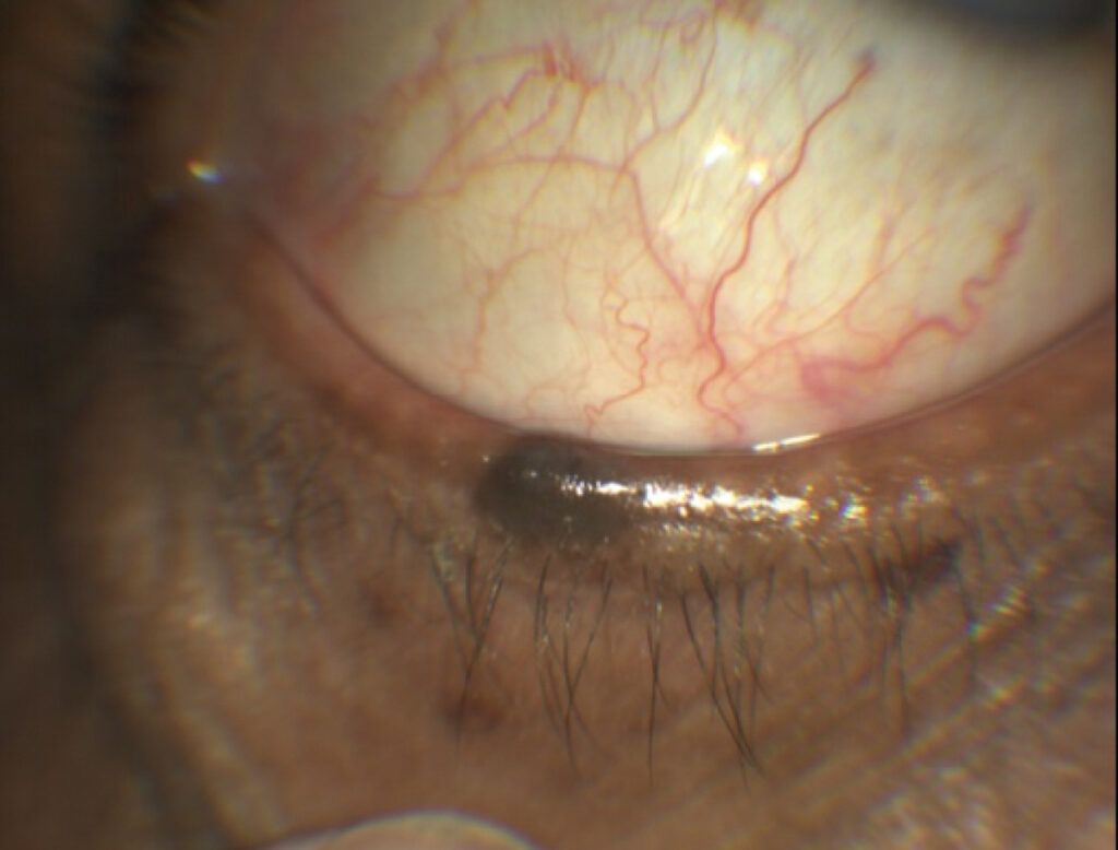
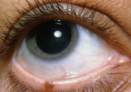
- A well-defined, round or oval lesion with a uniform brown color
- Smooth or slightly raised surface, sometimes with hair
- Stable size over years, or slow growth in childhood and adolescence
- A location on the lid margin or the skin, and sometimes extending to the conjunctiva
- Usually asymptomatic, with only cosmetic or irritative concern
A nevus on the lid margin may rub against the eye and cause irritation if it is large.
Features That Raise Concern
- Rapid enlargement or a change in shape
- Color change, including darkening, multiple colors, or loss of pigment
- Irregular borders and surface
- Ulceration, bleeding, or crusting
- Loss of eyelashes in the area (see madarosis)
- A new pigmented lesion in an adult
These features suggest atypical nevus or melanoma, and they call for biopsy.
Differential Diagnosis of Eyelid Nevus
- Seborrheic keratosis: a waxy, stuck-on lesion (see seborrheic keratosis of the eyelid)
- Pigmented basal cell carcinoma: a pearly lesion with telangiectasia (see basal cell carcinoma of the eyelid)
- Melanoma: rare on the eyelid but potentially serious
- Blue nevus and melanocytoma: deeper pigment with a blue-gray color
- Lentigo and freckles: flat pigmented spots from sun exposure
- Hemangioma and pyogenic granuloma: vascular lesions that can look dark
Management for Eyelid Nevus
Observation with photographs is reasonable for typical, stable nevi.
Also, measure the lesion and photograph it with a scale, and review at intervals.
Excise when there are suspicious features, when the lesion irritates the eye or the lashes, or for cosmetic reasons.
Send the specimen for histopathology, and ensure complete excision with margins, since pigmented lesions that are incompletely removed may recur or leave a confusing scar.
The method of excision depends on the site: shave excision, full-thickness pentagon excision, or staged reconstruction.
Counselling
Reassure the patient that most nevi are benign, and explain that a change in size, color, or shape is a reason to return.
Advise sun protection, since ultraviolet exposure contributes to the development of nevi and melanoma.
Arrange dermatologic examination of the rest of the skin if there are multiple or atypical nevi.
Dermoscopy and Imaging
Dermoscopy of the lid skin is feasible with a hand-held device.
It shows patterns such as regular globules, a reticular network, and homogeneous pigmentation in benign nevi.
Also, atypical features include irregular dots, streaks, and blue-white veil, and they raise the suspicion of melanoma.
Anterior segment imaging helps to see lesions on the lid margin and conjunctiva, and photographs allow comparison over time.
Excision Techniques
Small lid margin lesions may be excised with a shave technique that preserves the margin, and the tissue is sent for pathology.
In addition, larger lesions need a full-thickness pentagon excision, with repair of the margin, and large defects may require reconstruction with flaps.
In children with a divided nevus, the timing of surgery is individualized, since doctors must weigh the risk of amblyopia and the cosmetic effects.
When to Refer
Refer any lesion with a worrisome appearance or a pathology report that shows atypia or melanoma to an oculoplastic surgeon and a dermatologist or oncologist.
Also, patients with many atypical nevi or a family history of melanoma need a full skin examination at intervals.
For the latest studies, see the PubMed results for eyelid nevus.


Document what you see
Two smartphone imaging tools built for everyday clinic use — one for the slit lamp, one for the fundus.
From Choroida — the team behind this siteReferences
- Shields JA, Shields CL. Eyelid, Conjunctival, and Orbital Tumors: An Atlas and Textbook. 3rd ed. Wolters Kluwer; 2016.
- Cook BE Jr, Bartley GB. Epidemiologic characteristics and clinical course of patients with malignant eyelid tumors in an incidence cohort in Olmsted County, Minnesota. Ophthalmology. 1999;106:746-750.
- Salmon JF. Kanski’s Clinical Ophthalmology: A Systematic Approach. 9th ed. Elsevier; 2020.
- American Academy of Ophthalmology. Basic and Clinical Science Course, Section 7: Oculofacial Plastic and Orbital Surgery. AAO; current edition.

Writes and reviews the clinical articles on Educate. Every post is checked against peer-reviewed sources before it goes live.