OIS vs CRVO is a common clinical question because both conditions cause retinal hemorrhages, dilated veins and neovascular risk.
Ocular ischemic syndrome (OIS) reflects chronic arterial hypoperfusion, usually from severe carotid stenosis.
Central retinal vein occlusion (CRVO), however, reflects impaired venous outflow near the lamina cribrosa.
Therefore, a wrong label can delay carotid evaluation.
Why OIS vs CRVO Gets Confused
Both conditions affect older adults with hypertension, diabetes or other vascular risk factors.
Also, the term “venous stasis retinopathy” adds confusion (see venous stasis retinopathy).
Therefore, record the suspected mechanism in your notes.
Symptoms in OIS vs CRVO
CRVO usually causes sudden, painless blur.
In contrast, OIS tends to reduce vision over weeks or months.
Some OIS patients also report a dull ocular ache called ocular angina.
In addition, ask about transient visual loss.
Fundus Explorer Pro
Photograph the retinal findings described here with the phone already in your pocket — 22 D optics and built-in illumination in one handheld unit.
From Choroida — the team behind this siteFundus Findings in OIS vs CRVO
Hemorrhage pattern gives the first clue.
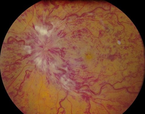
CRVO typically shows flame and blot hemorrhages in all four quadrants (see central retinal vein occlusion).
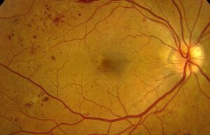
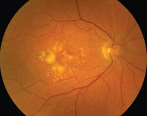
OIS usually shows fewer dot and blot hemorrhages in the midperiphery.
CRVO veins are dilated and markedly tortuous, while OIS veins are less tortuous.
Retinal arteries often look narrow in OIS.
Disc swelling favors acute CRVO, whereas the disc often looks normal in OIS.
Anterior Segment Clues
Iris neovascularization can complicate either disease, so examine the iris undilated and perform gonioscopy.
However, OIS may also produce flare and mild cells that mimic anterior uveitis.
For example, OIS eyes may show normal or low pressure despite angle neovascularization because ciliary body hypoperfusion reduces aqueous production.
Fluorescein Angiography in OIS vs CRVO
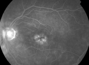
Delayed, patchy choroidal filling strongly suggests OIS.
OIS also commonly shows prolonged arteriovenous transit and late staining of arterioles.
In CRVO, angiography typically shows delayed venous filling, hemorrhagic blockage and variable capillary nonperfusion.
However, timing varies with injection technique and cardiac output, so interpret it clinically.
Reading Perfusion in Context
Peripheral nonperfusion occurs in both diseases, so it does not settle the diagnosis.
Widefield imaging can still map ischemia and guide laser planning.
Next, compare both eyes during the same session.
A normal fellow eye fits unilateral disease, whereas bilateral findings should prompt a broader review.
Electroretinography in OIS vs CRVO
Electroretinography is rarely required, but it can support a difficult diagnosis.
Ischemic CRVO often reduces the b-wave more than the a-wave.
OIS may reduce both waves because choroidal and retinal circulations can suffer.
Still, these patterns overlap, so they cannot replace vascular imaging.
Carotid Imaging
Order carotid duplex ultrasound when the examination suggests OIS.
Then consider CT or MR angiography when ultrasound is limited.
Also, a normal carotid study should prompt review of the diagnosis.
Systemic Clues in OIS vs CRVO
Prior stroke, transient ischemic attack or a carotid bruit increases suspicion for OIS.
However, a silent neck examination does not exclude severe stenosis.
CRVO is associated with hypertension, diabetes and glaucoma, so check all three.
Treatment Differences
CRVO care centers on macular edema and neovascular surveillance, commonly with anti-VEGF therapy.
The Central Vein Occlusion Study supported panretinal photocoagulation once iris neovascularization appears, but not prophylactic laser.
OIS management also treats neovascularization, but anti-VEGF benefit may be temporary (see OIS treatment options).
Neither approach corrects carotid stenosis.
Revascularization and Its Limits
Carotid endarterectomy aims mainly to lower stroke risk, and NASCET showed benefit in selected symptomatic high-grade stenosis.
Visual outcomes are less predictable, so avoid promising recovery.
In addition, restored perfusion may raise pressure when neovascularization has closed the angle.
Therefore, recheck pressure after the procedure.
Prognosis and Follow-Up
Poor initial acuity in CRVO predicts a higher risk of ischemic conversion.
Accordingly, schedule regular iris and angle checks early.
OIS carries a guarded visual prognosis once neovascular glaucoma develops.
Systemic prognosis also matters, since one older series reported five-year mortality near 40%, mostly cardiac.
Discussing OIS vs CRVO With Patients
Explain that a vein blockage and a narrowed neck artery can look similar inside the eye.
Next, explain why carotid imaging may be urgent.
Finally, stress that eye treatment cannot replace vascular risk management.
Pitfalls to Avoid
- Do not label asymmetric midperipheral hemorrhages as diabetic retinopathy
- Never let normal pressure reassure you when iris vessels are present
- Do not skip early angiographic frames, because choroidal filling may decide the diagnosis
- Never assume that treating the eye has addressed stroke risk


Document what you see
Two smartphone imaging tools built for everyday clinic use — one for the slit lamp, one for the fundus.
From Choroida — the team behind this siteReferences
- Brown GC, Magargal LE. The ocular ischemic syndrome: clinical, fluorescein angiographic and carotid angiographic features. Int Ophthalmol. 1988;11:239-251.
- Mizener JB, Podhajsky P, Hayreh SS. Ocular ischemic syndrome. Ophthalmology. 1997;104:859-864.
- Sivalingam A, Brown GC, Magargal LE, Menduke H. The ocular ischemic syndrome. II. Mortality and systemic morbidity. Int Ophthalmol. 1989;13:187-191.
- Central Vein Occlusion Study Group. Natural history and clinical management of central retinal vein occlusion. Arch Ophthalmol. 1997;115:486-491.
- North American Symptomatic Carotid Endarterectomy Trial Collaborators. Beneficial effect of carotid endarterectomy in symptomatic patients with high-grade carotid stenosis. N Engl J Med. 1991;325:445-453.

Writes and reviews the clinical articles on Educate. Every post is checked against peer-reviewed sources before it goes live.