Tear break-up time (TBUT) is the interval between a complete blink and the first appearance of a dry spot in the tear film.
It is the standard bedside measure of tear stability.
It is quick, inexpensive, and useful, but it is easy to perform badly, and a poorly done test gives numbers that mislead the diagnosis.
Understanding the technique and its limits makes the result more reliable.
What Is Tear Break-Up Time?
After a blink, the tear film spreads over the cornea and then thins and breaks up in a time that depends on the quality of the lipid layer, the mucin layer, and the surface.
A film that breaks up quickly leaves the epithelium exposed, which triggers discomfort, reflex tearing, and blurred vision.
Short break-up times are therefore a marker of tear film instability, which is the central problem in evaporative dry eye and in many mixed forms (see evaporative dry eye).
Clinicians use two versions: the invasive test with fluorescein, and non-invasive tests that use a reflected grid or a topographer to see the tear film without a dye.
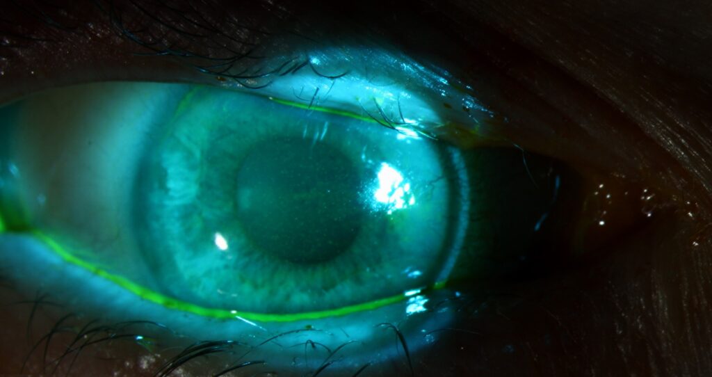
Tear Break-Up Time: Technique With Fluorescein
- Use a minimal volume of fluorescein, by wetting a strip with saline and shaking off the excess
- Touch the strip to the inferior conjunctiva without touching the cornea, and ask the patient to blink a few times
- Use a cobalt blue filter and a yellow barrier filter if available
- Ask the patient to blink once and then keep the eyes open, looking straight ahead
- Count the seconds from the blink to the first dark discontinuity
- Repeat the test two or three times and take the mean
The patient should not squeeze the lids, and the examiner should not touch the lids or the eye.
The room should be at a stable temperature and humidity, and the test should be done before other tests that disturb the tear film.
All-fit Slit-Lamp Adapter
Record and share exactly what you see at the slit lamp. One adapter fits any slit lamp or surgical microscope — and any smartphone.
From Choroida — the team behind this siteTear Break-Up Time: Interpretation
Clinicians generally consider values under 10 seconds abnormal, and values under 5 seconds suggest marked instability.
Normal fluorescein TBUT is often cited as above 10 seconds, but values vary across populations and methods.
Non-invasive break-up time is usually longer than fluorescein TBUT, because the dye itself destabilizes the film.
Clinicians also use a cut-off of about 10 seconds for it in diagnostic algorithms.
The pattern of break-up helps with interpretation.
- Area break: a large dry area, often in mucin-deficient states
- Line break: a streak along a line, associated with a tear meniscus or an irregular surface
- Spot break: a small round spot, often at a corneal irregularity
- Dimple break: small spots from lipid or surface tension effects
- Random break: a variable pattern
The location of the break gives a clue, since breaks at the same spot each time may reflect a surface irregularity such as a pterygium or a scar.
Common Pitfalls
Too much fluorescein stabilizes the tear film and falsely prolongs the time, while too little makes the break difficult to see.
Lid manipulation, a bright light, and a strong airflow can all shorten the time.
Reflex tearing from touching the eye or from a bright light can falsely lengthen it.
Different examiners count in different ways, so a single reading should not decide a diagnosis.
Use in Practice
The TBUT is one part of the diagnosis of dry eye disease, together with symptoms, osmolarity, and staining.
A short TBUT in a symptomatic patient with normal Schirmer values points to evaporative disease.
Treatment of the lid margin is the logical next step (see meibomian gland dysfunction).
Re-measure the TBUT after treatment, because an improvement gives the patient and the clinician a measure of progress.
Record the method and the value in the chart, since this makes comparison between visits meaningful.
Non-Invasive Break-Up Time
Non-invasive methods use a projected grid or ring and a topographer or keratograph to detect the first distortion of the reflected image without any dye.
The measurement is more reproducible and does not disturb the tear film, and many devices calculate the time automatically.
Its values are not interchangeable with fluorescein values, and the instrument manual should be consulted for the normal range.
Teaching Tip
When training a new examiner, have two people measure the same patient and compare, because the learning curve for judging the first break is short and the difference is instructive.
Standardize the volume of fluorescein, the lighting, and the number of repeats across the clinic.
Whichever method is used, record it in the chart in a consistent way, for example as the mean of three readings with the volume of fluorescein described.
For the latest studies, see the PubMed results for tear break-up time.


Document what you see
Two smartphone imaging tools built for everyday clinic use — one for the slit lamp, one for the fundus.
From Choroida — the team behind this siteReferences
- Wolffsohn JS, Arita R, Chalmers R, et al. TFOS DEWS II diagnostic methodology report. Ocul Surf. 2017;15:539-574.
- Cho P, Brown B. Review of the tear break-up time and a closer look at the tear break-up time of Hong Kong Chinese. Optom Vis Sci. 1993;70:30-38.
- Mannis MJ, Holland EJ, eds. Cornea. 4th ed. Elsevier; 2017.
- American Academy of Ophthalmology. Basic and Clinical Science Course, Section 8: External Disease and Cornea. AAO; current edition.

Writes and reviews the clinical articles on Educate. Every post is checked against peer-reviewed sources before it goes live.