Adaptive optics corrects the optical aberrations of the eye, and it allows imaging of individual photoreceptors in living patients.
Conventional fundus cameras and OCT cannot resolve cells because the cornea and lens blur the image.
Adaptive optics removes that blur, so researchers see the retinal mosaic directly.
How Adaptive Optics Works
A wavefront sensor measures the aberrations of the eye in real time.
A deformable mirror then changes its shape to cancel those aberrations, and a camera captures the corrected image.
Liang, Williams, and Miller reported the first retinal images with this approach in 1997.
Systems now combine adaptive optics with scanning laser ophthalmoscopy, flood illumination, or OCT.
What Adaptive Optics Shows
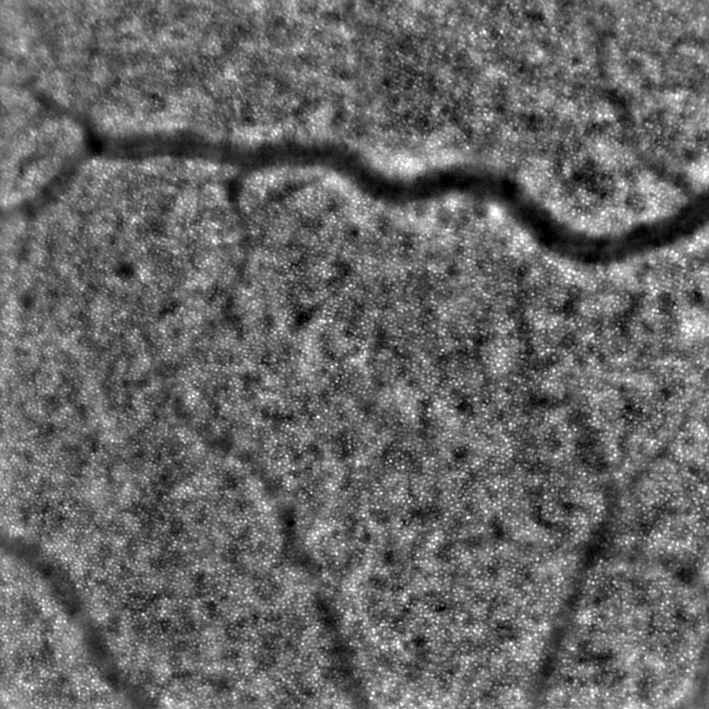
The cone mosaic appears as a regular array of bright spots, and rods and RPE cells appear in some systems.
Researchers also see microvessels, the nerve fiber layer, and the lamina cribrosa.
Measurements include cone density, spacing, and regularity, and these metrics change in disease (see OCT angiography).
Fundus Explorer Pro
Photograph the retinal findings described here with the phone already in your pocket — 22 D optics and built-in illumination in one handheld unit.
From Choroida — the team behind this siteClinical Uses of Adaptive Optics
- Inherited retinal diseases such as achromatopsia, Stargardt disease, and retinitis pigmentosa
- Monitoring of gene therapy and other trials
- Early detection of drug toxicity, such as hydroxychloroquine
- Study of vascular disease, including diabetic and hypertensive changes
In achromatopsia, for example, the images show a disrupted cone mosaic with a dark center (see achromatopsia).
Adaptive Optics in Trials
Gene therapy trials use cone counts and mosaic structure as outcome measures.
These measures may detect a response earlier than visual acuity or fields.
The technology also helps to prove that surviving cells remain after treatment.
Limits of Adaptive Optics
- High cost and limited availability
- A small field of view, often 1 to 3 degrees
- Need for a clear media and stable fixation
- Lack of normative databases and standard software
Image analysis requires specialized training, so many centers use adaptive optics for research only (see Stargardt disease).
Looking Ahead
Faster scanners and simpler systems may bring adaptive optics into clinics.
Automation will also reduce analysis time.
However, clinical use depends on proof that the findings improve decisions.
An Example Case
A 15-year-old boy has poor vision since birth, photophobia, and nystagmus, and the fundus looks nearly normal.
Genetic testing finds a mutation in a cone gene, consistent with achromatopsia.
Adaptive optics imaging shows a sparse cone mosaic with a dark center, while the rods appear intact.
The findings help the team to plan counseling, and they guide eligibility for a future gene therapy trial.
Questions Patients Ask
Patients ask whether adaptive optics is part of routine care, and the answer is that few centers offer it.
They also ask whether it will improve their vision, and the test itself gives information, not treatment.
Because the imaging is research based, consent and data use rules apply.
Pitfalls to Avoid
- Treating mosaic metrics as diagnostic without normative data
- Comparing images between devices without correction
- Overinterpreting small changes in cone density over time
- Forgetting that fixation errors can distort the mosaic
Looking Ahead
Compact systems may reach clinics, and automated analysis may speed up reporting.
Combined adaptive optics and OCT angiography may show cells and vessels together.
Therefore, expect broader use in trials of inherited disease and drug toxicity.
Practical Points
Refer patients to a research center when cell-level data may change management.
Then document the diagnosis and genetics, since centers often require them.
Also keep structural OCT and fundus autofluorescence results with the referral.
Technical Requirements
A system needs a stable head position, a dilated pupil, and good optical media.
Imaging sessions take longer than OCT, and the images need registration and averaging.
Because the field is small, operators choose locations of interest in advance.
Also expect training time for staff.
Interpreting Cone Metrics
Cone density falls with eccentricity and varies between individuals.
Therefore, compare the same eccentricity in follow-up images, and avoid conclusions from a single point.
Researchers also use spacing and regularity measures, which detect early disruption.
Finally, rods and cones differ in reflectivity, so identification needs careful software and expert review.
Normative data from the same device make these comparisons more reliable.
Share the images with the referring clinician, since the cone pattern often explains symptoms that standard tests cannot.
Then discuss the findings with the patient in simple terms, and explain what the images do and do not show.
Finally, keep copies in the research record with the genetic report.


Document what you see
Two smartphone imaging tools built for everyday clinic use — one for the slit lamp, one for the fundus.
From Choroida — the team behind this siteReferences
- Liang J, Williams DR, Miller DT. Supernormal vision and high-resolution retinal imaging through adaptive optics. J Opt Soc Am A. 1997;14:2884-2892.
- Roorda A, Duncan JL. Adaptive optics ophthalmoscopy. Annu Rev Vis Sci. 2015;1:19-50.
- Morgan JIW. The fundus photo has met its match: optical coherence tomography and adaptive optics ophthalmoscopy are here to stay. Ophthalmic Physiol Opt. 2016;36:218-239.
- American Academy of Ophthalmology. Basic and Clinical Science Course, Section 12: Retina and Vitreous. AAO; current edition.

Writes and reviews the clinical articles on Educate. Every post is checked against peer-reviewed sources before it goes live.