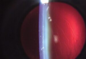
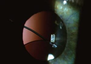
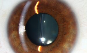
Scleral contact lens fitting uses large-diameter, rigid gas-permeable lenses that vault entirely over the cornea and rest instead on the more regular, less sensitive sclera.
That single design principle, resting on the sclera rather than the cornea, is what makes these lenses useful for eyes where a standard corneal lens either cannot achieve a stable fit or cannot be tolerated at all.
The reservoir of fluid trapped between the back of the lens and the front of the eye is doing real optical and therapeutic work, not just providing comfort.
Scleral lenses have grown well beyond their original niche in severe corneal disease, and they are now also used by some patients simply for the comfort and stability advantages over a standard soft or corneal rigid lens.
Design Principle
A scleral lens is large enough in diameter to clear the cornea entirely, creating a fluid-filled reservoir between the back surface of the lens and the front surface of the eye rather than the lens directly touching corneal tissue.
This fluid reservoir masks corneal irregularity optically, since the smooth anterior lens surface, not the irregular cornea underneath, becomes the eye’s effective refracting surface.
The lens vaults over the limbus and lands on the conjunctiva overlying the sclera, a comparatively insensitive and more uniformly curved surface that tolerates lens bearing far better than the cornea does.
Indications
- Irregular cornea from keratoconus, pellucid marginal degeneration, or other corneal ectasias, where a scleral lens’s smooth optical surface corrects irregular astigmatism that spectacles cannot
- Post-keratoplasty or post-refractive surgery corneas with irregular topography that a standard corneal lens cannot fit stably
- Severe ocular surface disease, including significant dry eye or exposure, where the fluid reservoir provides continuous corneal hydration and mechanical protection
- Corneal or limbal stem cell disease, where the lens shields a fragile ocular surface from the mechanical stress of blinking
All-fit Slit-Lamp Adapter
Record and share exactly what you see at the slit lamp. One adapter fits any slit lamp or surgical microscope — and any smartphone.
From Choroida — the team behind this siteHow the Fit Differs From Corneal Lenses
A corneal rigid gas-permeable or soft lens is fitted to align with and, to some degree, bear weight on the corneal surface itself, which becomes impossible once that surface is irregular enough to prevent a stable, centered fit.
A scleral lens sidesteps this problem entirely by never touching the cornea, so fitting focuses instead on achieving even, gentle landing on the conjunctiva and sclera, and confirming adequate corneal clearance across the entire vault.
Because the scleral surface itself is not perfectly spherical or symmetric between individuals, modern scleral lens fitting often benefits from corneal and scleral topography or profilometry to characterize the true shape being fitted, rather than relying on trial-and-error alone.
Toric or quadrant-specific peripheral curves are frequently needed to achieve an even landing across an asymmetric sclera, since a lens with a single uniform peripheral curve often lands unevenly and causes localized discomfort or compression.

Fitting Process and Follow-Up
Diagnostic lenses are placed and assessed with a slit lamp, checking central and peripheral corneal clearance, limbal clearance, and the evenness of scleral landing across all quadrants before settling on a final lens design.
Fluorescein, or optical coherence tomography in more advanced fitting practices, is used to visualize and quantify the fluid reservoir depth, since both too little clearance, risking corneal touch, and too much, risking suction and edema, are problems worth avoiding.
Follow-up after dispensing checks for signs of inadequate clearance, conjunctival compression from the landing zone, and midday fogging within the reservoir, adjusting the lens design as needed based on what is actually observed on wear.
Multiple fitting visits are typical rather than exceptional, and patients benefit from understanding upfront that arriving at a final, comfortable lens design is usually an iterative process rather than a single appointment.
Complications and Challenges
- Midday fogging, where debris accumulates in the tear reservoir over hours of wear, blurring vision and requiring the lens to be removed, cleaned, and reinserted
- Corneal hypoxia from restricted tear exchange under a large, sealed lens, mitigated by using highly oxygen-permeable materials and avoiding excessive vault
- Handling difficulty, since inserting and removing a large scleral lens filled with saline is considerably harder to learn than handling a standard soft contact lens
- Conjunctival compression or blanching from an uneven or overly tight landing zone, which can cause discomfort and, over time, localized conjunctival changes
Successful long-term wear depends heavily on patient education around insertion, removal, and cleaning technique, and practices that dedicate real time to hands-on training tend to see far better adherence and fewer complications than those that do not.


Document what you see
Two smartphone imaging tools built for everyday clinic use — one for the slit lamp, one for the fundus.
From Choroida — the team behind this siteReferences
- van der Worp E. A Guide to Scleral Lens Fitting.
- DeNaeyer G, Sanders DR, van der Worp E, et al. Qualitative assessment of scleral shape patterns using a new wide field ocular surface elevation topographer. Journal of Contact Lens Research and Science.
- American Academy of Ophthalmology. Basic and Clinical Science Course, Section 3: Clinical Optics.
Test yourself
A few questions straight from this article.
-
What defines a scleral contact lens?
Scleral lenses are large-diameter rigid gas-permeable lenses that clear the cornea entirely and land on the conjunctiva overlying the sclera. -
How does a scleral lens optically neutralise an irregular cornea?
The fluid reservoir masks corneal irregularity so that the smooth anterior lens surface, not the irregular cornea beneath, becomes the eye's effective refracting surface. -
Why is the sclera a better bearing surface than the cornea for a contact lens?
The lens lands on conjunctiva over the sclera, a comparatively insensitive and more uniformly curved surface that tolerates lens bearing far better than the cornea. -
Why does a scleral lens outperform spectacles in keratoconus?
In keratoconus and other ectasias the lens provides a smooth optical surface that corrects the irregular astigmatism spectacles are unable to address. -
What does the fluid reservoir provide in severe ocular surface disease?
In significant dry eye or exposure the reservoir keeps the cornea hydrated and shields a fragile surface from the mechanical stress of blinking. -
Compared with a corneal rigid lens, what does scleral lens fitting concentrate on?
Because the lens never touches the cornea, fitting targets gentle even landing on conjunctiva and sclera and confirms clearance across the whole vault. -
Why are toric or quadrant-specific peripheral curves often needed in scleral lens fitting?
The scleral surface is neither perfectly spherical nor symmetric, so a single uniform peripheral curve often lands unevenly and causes localised discomfort or compression. -
During scleral lens fitting, what risk comes from excessive corneal clearance?
Reservoir depth is assessed with fluorescein or OCT because too little clearance risks corneal touch while too much risks suction and edema. -
A scleral lens wearer reports vision blurring progressively through the afternoon, clearing after lens removal. What is this?
Debris accumulating in the reservoir over hours of wear blurs vision and requires the lens to be removed, cleaned and reinserted. -
How is corneal hypoxia under a sealed scleral lens minimised?
Restricted tear exchange under a large sealed lens risks hypoxia, mitigated by highly oxygen-permeable materials and by not vaulting the cornea excessively.

Writes and reviews the clinical articles on Educate. Every post is checked against peer-reviewed sources before it goes live.