
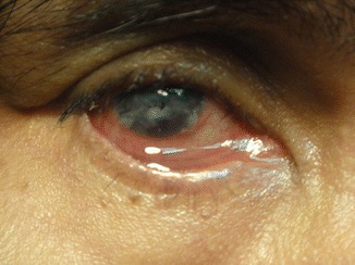
Exposure keratopathy is corneal surface damage caused by inadequate eyelid closure, leaving part or all of the cornea chronically unprotected from normal blinking and tear film coverage.
It is distinct from neurotrophic keratopathy, discussed in its own dedicated article on this site, where the primary defect is loss of corneal sensation rather than a mechanical failure of the lids to close, though the two can and often do coexist in the same patient.
Because the underlying cause is almost always identifiable and often treatable, exposure keratopathy is one of the more preventable causes of corneal scarring when it is recognized and addressed early.
It is also one of the more commonly overlooked diagnoses in a sedated or unconscious hospitalized patient, where incomplete eye closure is easy to miss amid other, more visible medical priorities.
Causes
- Facial nerve palsy, causing lagophthalmos from orbicularis oculi weakness, one of the most common causes encountered in general practice
- Thyroid eye disease, where proptosis and lid retraction together can prevent full lid closure despite normal facial nerve function
- Cicatricial lid disease, including ectropion or scarring from prior trauma, burns, or eyelid surgery, mechanically preventing normal apposition of the lids
- Reduced blink rate or incomplete blinking from a neurological or altered level of consciousness state, including sedation, anesthesia, or coma
- Proptosis of any cause, including orbital tumors or inflammatory disease, physically preventing the lids from closing over an eye pushed too far forward
Mechanism
Normal blinking spreads tears evenly across the cornea and periodically re-wets the surface, and normal lid closure during sleep protects the cornea from evaporation and mechanical exposure for hours at a stretch.
When lid closure is incomplete, whether from weakness, scarring, or mechanical obstruction, the exposed portion of cornea, most often the inferior third given gravity and typical lagophthalmos patterns, dries out and loses its protective tear film.
Chronic drying leads to epithelial breakdown, and if exposure continues without intervention, this can progress to corneal ulceration, scarring, and, in severe or neglected cases, perforation.
Secondary infection is a real risk once the epithelial barrier is compromised, since an exposed, drying cornea with an epithelial defect is considerably more vulnerable to microbial keratitis than an intact ocular surface.
All-fit Slit-Lamp Adapter
Record and share exactly what you see at the slit lamp. One adapter fits any slit lamp or surgical microscope — and any smartphone.
From Choroida — the team behind this siteClinical Presentation
Symptoms include foreign body sensation, redness, and blurred vision, often worse on waking if overnight lid closure is particularly inadequate.
Exam findings range from mild inferior punctate epithelial staining in early or mild cases to frank epithelial defects, corneal thinning, or infectious ulceration in more severe or longstanding exposure.
The degree of lagophthalmos, whether measured during a voluntary attempt at gentle closure or observed during sleep or sedation, correlates broadly with the severity of exposure, though even a small measurable gap can cause significant corneal disease over time if left unaddressed.
Evaluation
A careful assessment of lid closure, including measuring the degree of lagophthalmos and identifying the underlying cause, whether facial nerve palsy, thyroid eye disease, cicatricial change, or altered consciousness, guides both urgency and treatment approach.
Bell’s phenomenon, the normal reflexive upward rotation of the eye during attempted lid closure, is worth specifically assessing, since a poor or absent Bell’s phenomenon removes a natural protective mechanism and increases the risk of exposure damage when lid closure is incomplete.
Fluorescein staining characterizes the extent and severity of corneal surface damage and should be repeated at follow-up to track response to treatment.
Management
Frequent lubrication with artificial tears during the day and a thicker ointment at night forms the foundation of treatment for mild to moderate cases, aiming to replace the protective function normal blinking and lid closure would otherwise provide.
Taping the lids closed at night, or using a moisture chamber, can provide additional protection in patients with significant nocturnal lagophthalmos, particularly in an intensive care or perioperative setting where the cause is temporary.
Addressing the underlying cause directly, whether facial nerve palsy treatment, thyroid eye disease management, or eyelid reconstructive surgery for cicatricial disease, is the definitive treatment for cases where medical management alone is insufficient or the underlying process is not expected to resolve on its own.
A temporary or permanent tarsorrhaphy, partially suturing the lids together, is reserved for severe or refractory exposure that has not responded to lubrication and treatment of the underlying cause, providing mechanical protection while a more definitive plan is pursued.
In an inpatient setting, simple, low-cost preventive measures like regular ointment application and lid taping for a sedated patient are worth building into routine nursing protocols, since the resulting corneal damage is otherwise entirely avoidable.


Document what you see
Two smartphone imaging tools built for everyday clinic use — one for the slit lamp, one for the fundus.
From Choroida — the team behind this siteReferences
- Latkany RL, Lock B, Speaker M. Nocturnal lagophthalmos: an overview and classification. The Ocular Surface.
- American Academy of Ophthalmology. Basic and Clinical Science Course, Section 8: External Disease and Cornea.
- Bowling B. Kanski’s Clinical Ophthalmology.
Test yourself
A few questions straight from this article.
-
What causes the corneal damage in exposure keratopathy?
Incomplete lid closure leaves part or all of the cornea chronically unprotected from normal blinking and tear film coverage. -
Which feature separates neurotrophic keratopathy from exposure keratopathy?
In neurotrophic keratopathy the problem is loss of corneal sensation rather than a mechanical failure of the lids to close, although the two often coexist. -
Weakness of which muscle produces lagophthalmos in facial nerve palsy?
Facial nerve palsy weakens the orbicularis oculi, and the resulting lagophthalmos is one of the commonest causes seen in general practice. -
Why does thyroid eye disease prevent full lid closure?
Proptosis and lid retraction together can stop the lids meeting even though facial nerve function is entirely normal. -
Which part of the cornea is most often affected in exposure keratopathy?
Given gravity and the usual pattern of lagophthalmos, it is most often the inferior third of the cornea that dries and loses its tear film. -
What does Bell's phenomenon describe?
Bell's phenomenon is the normal reflex upward rotation of the eye during attempted lid closure, and a poor or absent response removes a natural protective mechanism. -
What added risk follows breakdown of the corneal epithelial barrier in exposure keratopathy?
An exposed, drying cornea with an epithelial defect is considerably more vulnerable to microbial keratitis than an intact ocular surface. -
What forms the foundation of treatment in mild to moderate exposure keratopathy?
Frequent lubrication by day and a thicker ointment overnight replaces the protection that normal blinking and lid closure would otherwise give. -
Which measure protects an eye with significant nocturnal lagophthalmos?
Lid taping or a moisture chamber adds protection overnight, and is especially useful in intensive care or perioperative settings where the cause is temporary. -
When is tarsorrhaphy used in exposure keratopathy?
Partially suturing the lids is reserved for severe or refractory exposure that has not responded to lubrication and treatment of the underlying cause.

Writes and reviews the clinical articles on Educate. Every post is checked against peer-reviewed sources before it goes live.


