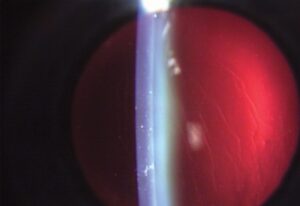
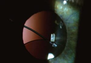
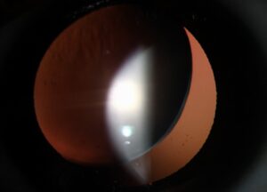
Refractive lens exchange (RLE) is the removal of a clear or early cataractous lens and its replacement with an intraocular lens to correct refractive error and presbyopia.
The operation is identical in technique to cataract surgery, but the patient has a different set of expectations and risks.
The challenge for the surgeon is to choose patients wisely, because the downsides are greater than for laser procedures in some groups.
What Is Refractive Lens Exchange?
Clinicians use RLE when laser vision correction is unsuitable, for example in high hyperopia, in presbyopes who want less dependence on glasses, and in some eyes with thin corneas.
Replacing the lens eliminates future cataract, and a multifocal, extended depth of focus, or accommodating lens gives a range of vision.
Patients with high myopia are poor candidates, because the risk of retinal detachment is higher.
The age at which RLE is performed also matters: the procedure sacrifices the remaining accommodation in a younger patient, so patients over 45 or 50 are better candidates.
Refractive Lens Exchange: Candidate Selection
- Presbyopic patients, usually over 45 to 50, who want to reduce the need for reading glasses
- High hyperopia, which is difficult to treat with laser (see presbyopia)
- Early lens changes in a patient with a refractive problem
- Corneas that are too thin or irregular for laser surgery
- A healthy macula and optic nerve, with realistic expectations
Relative contraindications include a retinal predisposition, such as lattice degeneration with holes, a history of retinal detachment, uveitis, macular disease, severe dry eye, and zonular weakness.
All-fit Slit-Lamp Adapter
Record and share exactly what you see at the slit lamp. One adapter fits any slit lamp or surgical microscope — and any smartphone.
From Choroida — the team behind this siteRisks of Refractive Lens Exchange
- Retinal detachment: the most serious risk, higher in myopic and younger patients and after posterior capsule rupture (see pseudophakic retinal detachment)
- Endophthalmitis: a rare but devastating infection (see acute postoperative endophthalmitis)
- Cystoid macular edema (see Irvine-Gass syndrome)
- Posterior capsule opacification (see YAG laser capsulotomy)
- Refractive surprise, especially in eyes with unusual biometry
- Dysphotopsia and loss of contrast with multifocal lenses (see negative dysphotopsia)
- Loss of accommodation in younger patients
The long-term retinal detachment risk is greater in axially long eyes than in short eyes.
The data suggest that it is higher than after laser vision correction.
Preoperative Evaluation
Take a careful history of refractive stability and expectations.
Perform a complete examination with a dilated fundus view, including the periphery, and OCT of the macula.
Use biometry with modern formulas, corneal topography, and an assessment of the tear film.
Warn the patient about the possible need for additional procedures such as laser enhancement.
Counselling
Explain that RLE is a lens operation with the same risks as cataract surgery, and that the surgery is done in an eye that has no cataract.
Discuss the alternatives, and the compromises of multifocal lenses, including halos and reduced contrast, particularly at night.
Obtain realistic expectations: a high degree of spectacle independence is likely but not guaranteed.
Outcomes of Refractive Lens Exchange
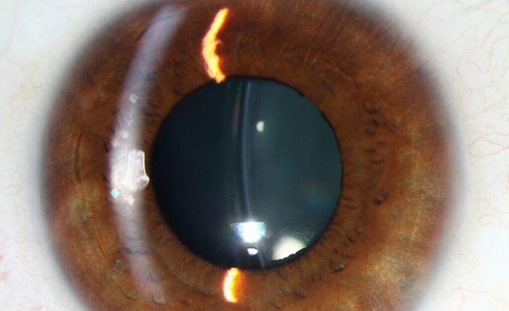
Visual acuity and spectacle independence are high in well-selected patients, and satisfaction is good when expectations match the result.
Patients who are dissatisfied typically had unrealistic expectations, ocular surface disease, or residual refractive error.
Choosing the Lens
The choice depends on the patient’s priorities and the health of the eye: a monofocal lens for the best contrast, a multifocal or extended depth of focus lens for spectacle independence, or a toric lens to correct corneal astigmatism.
Multifocal lenses carry a higher risk of halos and glare, so they suit patients who are highly motivated and who have healthy eyes, with no macular disease, glaucoma, or ocular surface disease.
A blended approach, with different lenses in the two eyes, is sometimes used.
Biometry and Formulas
Short and long eyes are at higher risk of refractive surprise.
A modern formula that accounts for axial length and lens position gives better accuracy.
Eyes that have had refractive surgery need special formulas (see IOL power after refractive surgery).
Keep the target in mind, since many surgeons aim for a mild minus in the non-dominant eye to improve near vision.
Retinal Risk Management
Every candidate should have a dilated peripheral fundus examination before surgery, with prophylactic treatment of any suspicious lesions.
Surgeons should avoid posterior capsule rupture and vitreous loss, which raise the risk of detachment.
Tell patients the warning symptoms.
A patient with prior retinal tears or lattice needs an individual risk discussion, and in some cases an alternative approach is safer.
When RLE Is Not the Best Choice
For young myopes with healthy corneas, laser vision correction or an implantable collamer lens may carry less risk to the retina.
Patients with unrealistic expectations or active ocular surface disease should be treated first or counselled against surgery.
For the latest studies, see the PubMed results for refractive lens exchange.


Document what you see
Two smartphone imaging tools built for everyday clinic use — one for the slit lamp, one for the fundus.
From Choroida — the team behind this siteReferences
- Alió JL, Grzybowski A, Romaniuk D. Refractive lens exchange in modern practice: when and when not to do it? Eye Vis (Lond). 2014;1:10.
- Ripandelli G, Billi B, Fedeli R, Stirpe M. Retinal detachment after clear lens extraction in 41 eyes with high axial myopia. Retina. 1996;16:3-6.
- Salmon JF. Kanski’s Clinical Ophthalmology: A Systematic Approach. 9th ed. Elsevier; 2020.
- American Academy of Ophthalmology. Basic and Clinical Science Course, Section 13: Refractive Surgery. AAO; current edition.

Writes and reviews the clinical articles on Educate. Every post is checked against peer-reviewed sources before it goes live.