The AREDS simplified severity scale estimates the five-year risk of advanced AMD from two fundus findings.
Also, clinicians can apply it at the slit lamp or on photographs without special software.
Therefore, it turns a vague impression of early disease into a number that guides follow-up and supplements.
How the AREDS Simplified Severity Scale Works
Ferris and colleagues derived the scale from the Age-Related Eye Disease Study.
First, two risk factors count in each eye: large drusen and pigmentary abnormalities.
The score is the sum of both eyes, from 0 to 4, and each step carries a higher risk (see what is geographic atrophy).
Definitions Used in the AREDS Simplified Severity Scale
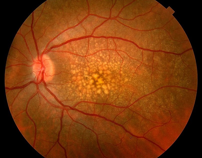
- Large drusen: at least one druse of 125 micrometers or more, about the width of a retinal vein at the disc margin
- Pigmentary abnormalities: increased pigment or depigmentation related to AMD in the inner subfield of the macula
- Eyes with advanced AMD count as 2 in the score
One point is assigned for each factor in each eye, and a person with advanced AMD in one eye receives credit as if both factors were present.
Fundus Explorer Pro
Photograph the retinal findings described here with the phone already in your pocket — 22 D optics and built-in illumination in one handheld unit.
From Choroida — the team behind this siteFive-Year Risk With the AREDS Simplified Severity Scale
The risk of advanced AMD in at least one eye rises steeply with the score.
- Zero points mean about 0.5 percent risk
- One point raises the risk to about 3 percent
- With two points, risk reaches about 12 percent
- Three points correspond to about 25 percent
- A full score of 4 predicts about 50 percent
However, these values come from the AREDS cohort and may differ in other populations.
Using the AREDS Simplified Severity Scale in Practice
Because patients with scores of 0 or 1 have a low risk, they need routine follow-up.
In contrast, patients with higher scores deserve counseling on home monitoring and closer visits.
The AREDS2 formulation reduces progression in intermediate disease, so consider it for patients in the higher categories (see areds supplements and AMD).
Strengths and Limits of the AREDS Simplified Severity Scale
Also, the scale is simple and validated in a large trial.
However, it does not include OCT features such as hyperreflective foci or subretinal drusenoid deposits.
Newer models add imaging and genetics, but the scale remains useful for quick counseling.
An Example Case
A 68-year-old woman has several large drusen in the right eye and a patch of pigment clumping in the left eye.
She scores 1 for large drusen in the right eye, and 1 for pigment and 1 for large drusen in the left eye, so the total is 3.
The clinician estimates a five-year risk near 25 percent for advanced AMD in one eye.
She starts the AREDS2 supplement, uses an Amsler grid each day, and returns every six months.
Questions Patients Ask
Patients ask what the number means, and it is a group risk, not a prediction for their eyes.
They also ask whether the risk can be lowered, and supplements, no smoking, and a healthy diet may help.
Because numbers can frighten, present the score along with a plan.
Pitfalls to Avoid
- Counting small drusen, which do not add to the score
- Missing pigmentary change in the inner subfield
- Applying the scale to eyes with advanced disease without adjusting
- Forgetting the fellow eye when estimating risk
Looking Ahead
Newer models combine OCT markers, genetics, and clinical data to improve risk estimates.
Also, deep learning may grade fundus photographs automatically.
Therefore, the simple scale may be replaced by automated scores that follow the same logic.
Practical Points
Grade each eye on color photographs or at the slit lamp with a dilated pupil.
Then record the score and the date in the chart.
Also plan follow-up from the score, with closer visits for scores 3 and 4.
From Score to Plan
For scores 0 and 1, review every one to two years.
For scores 2 and above, consider supplements and home monitoring.
Because risk rises steeply, shorten the interval at higher scores.
What the AREDS Simplified Severity Scale Misses
The scale ignores reticular pseudodrusen, hyperreflective foci, and other OCT features.
Eyes with these findings may be at higher risk than the score suggests.
Therefore, interpret the number together with imaging.
Recording the Score
Write the score for each eye and the total in the chart.
Then add the date and the image used for grading.
Also repeat the grading yearly, since drusen and pigment change over time.


Document what you see
Two smartphone imaging tools built for everyday clinic use — one for the slit lamp, one for the fundus.
From Choroida — the team behind this siteReferences
- Ferris FL, Davis MD, Clemons TE, et al. A simplified severity scale for age-related macular degeneration: AREDS report no. 18. Arch Ophthalmol. 2005;123:1570-1574.
- Davis MD, Gangnon RE, Lee LY, et al. The Age-Related Eye Disease Study severity scale for age-related macular degeneration: AREDS report no. 17. Arch Ophthalmol. 2005;123:1484-1498.
- AREDS Research Group. A randomized, placebo-controlled, clinical trial of high-dose supplementation with vitamins C and E, beta carotene, and zinc for age-related macular degeneration and vision loss: AREDS report no. 8. Arch Ophthalmol. 2001;119:1417-1436.
- Chew EY, Clemons TE, Sangiovanni JP, et al. Lutein + zeaxanthin and omega-3 fatty acids for age-related macular degeneration: the AREDS2 randomized clinical trial. JAMA. 2013;309:2005-2015.

Writes and reviews the clinical articles on Educate. Every post is checked against peer-reviewed sources before it goes live.