
Corneal melt, or sterile keratolysis, is progressive stromal thinning driven by excessive collagenase and other matrix-degrading enzyme activity, occurring without active microbial infection, distinguishing it clearly from an infectious corneal ulcer.
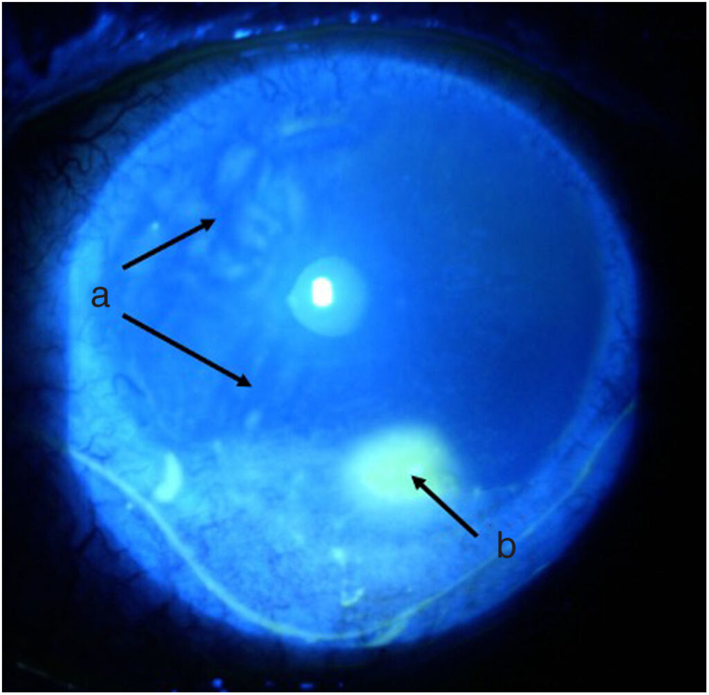
The distinction from infectious ulceration matters enormously for treatment, since a sterile melt often requires controlling inflammation and matrix breakdown, sometimes including corticosteroids, while an infectious ulcer demands antimicrobial therapy and can be actively worsened by steroid use before infection is controlled.
Understanding the range of underlying causes, from systemic autoimmune disease to a purely local postoperative or exposure-related process, is essential to identifying and addressing the actual driver behind any individual case of corneal melt.
A confirmed corneal melt should always prompt the question of whether it represents purely local disease or a window into previously unrecognized systemic illness, since the answer changes the entire management plan.
Mechanism
Corneal stromal integrity depends on a normal balance between collagen synthesis and the activity of matrix metalloproteinases and other enzymes that break collagen down as part of ordinary tissue turnover and remodeling.
In corneal melt, this balance shifts decisively toward degradation, whether from excessive enzyme production, insufficient enzyme inhibition, or both, driven by an underlying inflammatory, autoimmune, or severe surface disease process.
Because no active microbial infection is driving this process, standard antimicrobial therapy alone does not address the actual mechanism, which is why correctly identifying a sterile melt as sterile, rather than assuming it must be infectious, has such direct treatment implications.
Causes
- Rheumatoid arthritis and other systemic autoimmune connective tissue diseases, among the most classic and well-recognized causes of peripheral ulcerative keratitis and corneal melt, discussed in relation to peripheral ulcerative keratitis elsewhere on this site
- Severe dry eye disease or exposure keratopathy, discussed in its own dedicated article on this site, where chronic surface compromise predisposes to progressive stromal breakdown
- Neurotrophic keratopathy, discussed in its own dedicated article on this site, where impaired corneal sensation and healing capacity predispose to progressive thinning
- Postoperative or post-infectious melt, occurring after ocular surgery or following resolution of an infectious keratitis, where residual inflammation continues driving matrix breakdown even after the original insult has resolved
- Topical medication toxicity, including from preservatives in chronic topical drop use or from certain classes of topical anti-inflammatory medication used excessively
All-fit Slit-Lamp Adapter
Record and share exactly what you see at the slit lamp. One adapter fits any slit lamp or surgical microscope — and any smartphone.
From Choroida — the team behind this siteClinical Presentation
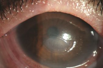
Progressive stromal thinning, often at the peripheral or paracentral cornea, presents with variable pain, redness, and vision changes depending on the location and rate of thinning, sometimes surprisingly mild given how alarming the thinning itself can appear on exam.
Advanced cases can progress to descemetocele formation, discussed in its own dedicated article on this site, or frank corneal perforation if the underlying process is not identified and controlled promptly.
The absence of a significant infiltrate or the typical clinical picture of active infection, combined with a relevant systemic or local history, should raise suspicion for a sterile process rather than infectious keratitis, though the two can sometimes coexist or be difficult to distinguish with certainty on clinical grounds alone.
Evaluation
A thorough systemic review, including screening for rheumatoid arthritis and other autoimmune connective tissue disease, is essential whenever corneal melt is identified without an obvious local explanation, given how often it can be the first presenting sign of previously undiagnosed systemic disease.
Culture and, when infection cannot be confidently excluded clinically, empiric coverage for possible infectious keratitis are appropriate initial steps, since missing an actual infection while treating presumed sterile melt with immunosuppressive therapy could be genuinely dangerous.
A careful review of the ocular surface, tear film, and any current or recent topical medications helps identify local contributing factors that need to be addressed alongside any systemic disease found on workup.
Management
Treating any identified underlying systemic disease, in coordination with rheumatology when autoimmune disease is confirmed, is essential, since local ocular treatment alone often fails to halt progression when a systemic inflammatory driver remains uncontrolled.
Topical and, in significant cases, oral matrix metalloproteinase inhibitors, along with aggressive lubrication and, once infection has been reasonably excluded, topical corticosteroids, are used to control local inflammation and enzymatic breakdown.
Tectonic support, including a bandage contact lens, tissue adhesive, amniotic membrane transplantation, discussed in its own dedicated article on this site, or in severe cases a corneal patch graft, may be needed to prevent or address perforation while the underlying process is brought under control.
Close, frequent monitoring during the active phase is essential regardless of which specific treatments are chosen, since the corneal thickness can change meaningfully over just days when the underlying enzymatic process is still actively progressing.


Document what you see
Two smartphone imaging tools built for everyday clinic use — one for the slit lamp, one for the fundus.
From Choroida — the team behind this siteReferences
- Messmer EM, Foster CS. Vasculitic peripheral ulcerative keratitis. Survey of Ophthalmology.
- American Academy of Ophthalmology. Basic and Clinical Science Course, Section 8: External Disease and Cornea.
- Yagci A. Update on peripheral ulcerative keratitis. Clinical Ophthalmology.
Test yourself
A few questions straight from this article.
-
What characterises corneal melt, also called sterile keratolysis?
Corneal melt is progressive stromal thinning driven by excessive collagenase and other matrix-degrading enzyme activity, occurring without active microbial infection. -
Which imbalance underlies the stromal loss in corneal melt?
Stromal integrity depends on balance between collagen synthesis and matrix metalloproteinase activity. In melt this balance shifts decisively toward degradation, from excess enzyme, insufficient inhibition, or both. -
Why does distinguishing sterile melt from infectious ulceration change management so sharply?
Sterile melt often needs inflammation and matrix breakdown controlled, sometimes with corticosteroids, whereas an infectious ulcer needs antimicrobials and can be actively worsened by steroids before the infection is controlled. -
Which systemic disease is among the most classic causes of corneal melt?
Rheumatoid arthritis and other systemic autoimmune connective tissue diseases are among the most classic and well-recognised causes of peripheral ulcerative keratitis and corneal melt. -
Which examination feature supports a sterile rather than infectious cause of corneal thinning?
Lack of a significant infiltrate or the typical picture of active infection, together with a relevant systemic or local history, points toward a sterile process, though the two can coexist. -
What can advanced, uncontrolled corneal melt progress to?
If the underlying process is not identified and controlled promptly, advanced cases can progress to descemetocele formation or frank corneal perforation. -
Which evaluation step is essential in corneal melt without an obvious local explanation?
Corneal melt can be the first presenting sign of previously undiagnosed systemic disease, so a thorough systemic review including autoimmune connective tissue screening is essential. -
Why is culture or empiric antimicrobial cover appropriate before treating a presumed sterile melt?
When infection cannot be confidently excluded clinically, culture and empiric cover are appropriate, because treating a genuine infection with immunosuppressive therapy instead could be genuinely dangerous. -
Which measure provides tectonic support for a thinning cornea at risk of perforation?
Tectonic support options include a bandage contact lens, tissue adhesive, amniotic membrane transplantation, or in severe cases a corneal patch graft, used while the underlying process is controlled. -
Why does active corneal melt require close, frequent monitoring?
While the enzymatic process is still actively progressing, corneal thickness can change meaningfully over just days, so frequent review during the active phase is essential.

Writes and reviews the clinical articles on Educate. Every post is checked against peer-reviewed sources before it goes live.


