Vitreous haze grading estimates how much vitreous inflammation obscures the view of the fundus.
The score uses the optic disc and retinal vessels as landmarks.
It therefore gives uveitis clinicians a common language for activity and treatment response.
What Vitreous Haze Grading Measures
Haze reflects protein and cellular debris scattering light within the vitreous.
Therefore, the examiner judges fundus clarity and does not count individual cells.
Symptoms often track the score poorly.
Macular edema, for example, can reduce acuity while haze stays mild.
Origin of the NEI Scale
Nussenblatt and colleagues published the National Eye Institute scale in 1985.
They matched clinical views with standard photographs, using grades from 0 to 4+.
Later, the SUN Working Group endorsed it for reporting uveitis activity (see SUN classification).
Fundus Explorer Pro
Photograph the retinal findings described here with the phone already in your pocket — 22 D optics and built-in illumination in one handheld unit.
From Choroida — the team behind this siteVitreous Haze Grading Steps
Published wording varies slightly, but these descriptions reflect common clinical use:
- 0: clear view, with no haze
- Trace: slight blurring of the disc margin, with loss of the normal nerve fiber layer reflex
- 1+: mild blurring of the disc and retinal vessels
- 2+: moderate blurring of the disc and vessels
- 3+: disc visible, but its borders are markedly blurred and vessel detail is poor
- 4+: disc not visible
Slit Lamp Technique for Vitreous Haze Grading
First, dilate the pupil fully, because a small pupil dims the view and inflates the score.
Also, record lens status, since cataract can mimic haze.
Then use a 78 D or 90 D lens and center the disc (see slit lamp illumination).
Keep illumination moderate, because a bright beam can make a hazy view look clearer.
However, the original standards relied on indirect ophthalmoscopy, so confirm uncertain scores with that view.
Haze Versus Vitreous Cells
Cells appear as discrete particles in the anterior vitreous, while haze blurs the entire view.
The two findings can diverge in the same eye.
For example, cells may persist after haze clears in chronic disease.
Therefore, record them separately.
Media That Confound Vitreous Haze Grading
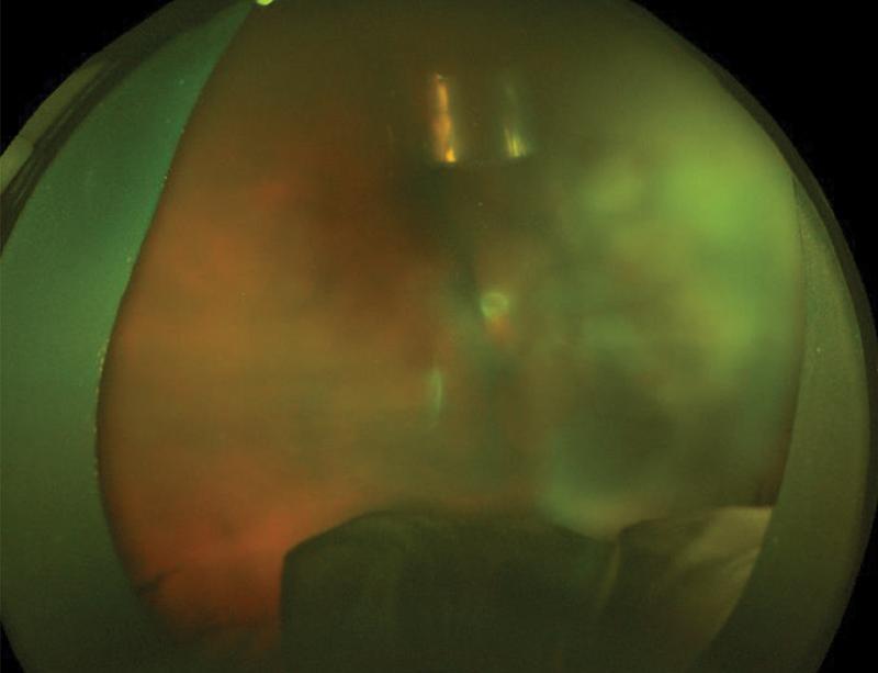
Corneal edema, posterior synechiae, cataract and capsule opacity all reduce fundus clarity.
As a result, compare the view with the fellow eye and document the limiting medium.
Vitreous hemorrhage and asteroid hyalosis can also mimic inflammatory haze.
In addition, consider lymphoma in older patients with steroid-resistant opacity.
Infection must also be excluded when haze rises rapidly after surgery or injection.
Reliability of Vitreous Haze Grading
Clinical scoring remains subjective, and experienced examiners can disagree.
Kempen and colleagues found limited interobserver agreement among uveitis specialists.
Therefore, interpret one-step differences cautiously.
When possible, the same clinician should repeat the examination.
Photographic Scales and OCT
Davis and colleagues developed a nine-step photographic scale for finer measurement.
However, it needs good images and trained readers, so it fits trials better than routine clinics.
Keane and colleagues showed that OCT vitreous signal correlated with clinical haze scores.
Still, it needs further validation, although OCT also reveals macular edema.
Using Vitreous Haze Grading in Treatment Decisions
SUN defines worsening as a two-step rise in activity, or a move from 3+ to 4+.
Similarly, improvement means a two-step fall, or a fall to grade 0.
These definitions serve reporting, so they do not dictate treatment alone.
Instead, weigh vision threat, macular edema, vasculitis, systemic disease and previous response.
For example, 1+ haze with macular edema may need treatment, while stable trace haze may not.
Options include local corticosteroid, systemic corticosteroid and steroid-sparing therapy, once infection is excluded.
VISUAL I also included vitreous haze in its treatment failure criteria.
Vitreous Haze Grading in Follow-Up
Record the grade, method, lens status and fellow eye findings at each visit.
Serial fundus photographs can also help when several clinicians share the care.
During a steroid taper, recheck haze along with acuity and OCT.
If haze persists without other activity, consider inactive debris before escalating immunosuppression.
Also, warn patients that floaters may persist even when inflammation improves.
Ask them to report pain, redness or reduced vision promptly.
Pitfalls to Avoid
- Do not grade through an undilated pupil
- Do not treat a one-step change as definite progression
- Avoid attributing cataract-related blur to vitreous inflammation
- Do not let a low grade reassure you when macular edema is present


Document what you see
Two smartphone imaging tools built for everyday clinic use — one for the slit lamp, one for the fundus.
From Choroida — the team behind this siteReferences
- Nussenblatt RB, Palestine AG, Chan CC, Roberge F. Standardization of vitreal inflammatory activity in intermediate and posterior uveitis. Ophthalmology. 1985;92:467-471.
- Jabs DA, Nussenblatt RB, Rosenbaum JT. Standardization of uveitis nomenclature for reporting clinical data. Am J Ophthalmol. 2005;140:509-516.
- Davis JL, Madow B, Cornejo J, et al. Scale for photographic grading of vitreous haze in uveitis. Am J Ophthalmol. 2010;150:637-641.
- Kempen JH, Ganesh SK, Sangwan VS, Rathinam SR. Interobserver agreement in grading activity and site of inflammation in eyes of patients with uveitis. Am J Ophthalmol. 2008;146:813-818.
- Keane PA, Karampelas M, Sim DA, et al. Objective measurement of vitreous inflammation using optical coherence tomography. Ophthalmology. 2014;121:1706-1714.
- Jaffe GJ, Dick AD, Brezin AP, et al. Adalimumab in patients with active noninfectious uveitis. N Engl J Med. 2016;375:932-943.

Writes and reviews the clinical articles on Educate. Every post is checked against peer-reviewed sources before it goes live.