
Limbal stem cell deficiency (LSCD) occurs when the stem cell population at the corneal limbus that normally renews and maintains a clear corneal epithelium is lost or dysfunctional, allowing conjunctival tissue to grow across the corneal surface in its place.
The result is a cornea that cannot repair itself normally, with vascularization, chronic epithelial defects, and gradually worsening vision that does not respond to the usual treatments for ocular surface disease.
Why Limbal Stem Cells Matter
The limbus, the ring of tissue at the corneal-conjunctival border, houses the stem cells responsible for continuously renewing the corneal epithelium and acting as a barrier that keeps conjunctival tissue from migrating onto the cornea.
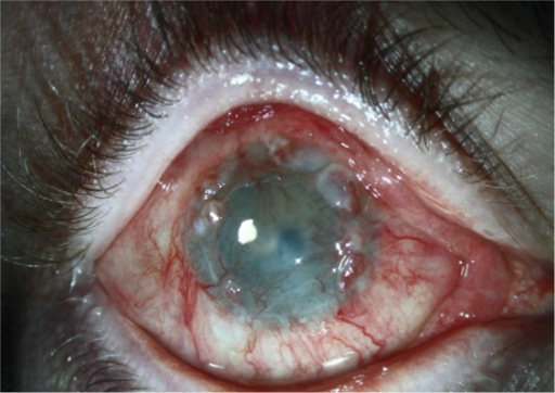
When this population is destroyed or overwhelmed, the cornea loses both its capacity for normal epithelial turnover and its barrier against conjunctival overgrowth, a process called conjunctivalization, in which vascularized, goblet-cell-containing conjunctival epithelium replaces the normally avascular corneal surface.
Causes
Acquired
- Chemical or thermal burns, among the most common and severe causes
- Stevens-Johnson syndrome and toxic epidermal necrolysis (see Stevens-Johnson syndrome ocular sequelae)
- Ocular cicatricial pemphigoid
- Contact lens-related chronic hypoxia and toxicity, usually milder and more localized
- Multiple ocular surface surgeries or chronic limbal inflammation
- Severe microbial keratitis involving the limbus
Congenital
- Aniridia, in which limbal stem cell deficiency develops progressively over childhood and adolescence (see aniridia-related keratopathy)
- Congenital ectodermal dysplasia syndromes
All-fit Slit-Lamp Adapter
Record and share exactly what you see at the slit lamp. One adapter fits any slit lamp or surgical microscope — and any smartphone.
From Choroida — the team behind this siteClinical Features
- Conjunctivalization, seen as a dull, irregular, vascularized epithelium crossing onto the cornea
- Persistent or recurrent epithelial defects
- Chronic inflammation and superficial neovascularization
- Reduced vision from surface irregularity, scarring, and vascularization
- Late fibrovascular pannus in advanced disease
Disease can be focal, sectoral, or total, and unilateral or bilateral, which affects both prognosis and treatment options.
Diagnosis
The diagnosis is largely clinical, based on the characteristic appearance of conjunctivalized epithelium on the cornea, often confirmed with impression cytology showing goblet cells on the corneal surface, which are normally restricted to the conjunctiva.
In vivo confocal microscopy can assess residual limbal stem cell architecture and support the diagnosis in equivocal cases.
Staging by the extent of limbal involvement, from focal sectoral disease to total 360-degree involvement, guides the choice of treatment.
Management
Medical and Supportive Care
Aggressive lubrication, treatment of any underlying inflammatory disease, and punctal occlusion support the ocular surface in mild or partial disease.
Autologous serum eye drops supply growth factors that support epithelial health in some patients.
Surgical Options
- Conjunctival epitheliectomy alone for very focal disease, removing the encroaching tissue
- Limbal stem cell transplantation, using tissue from the fellow eye when disease is unilateral, from a living relative, or from a cadaver donor, combined with systemic immunosuppression for allografts
- Cultivated limbal or oral mucosal epithelial transplantation, an evolving technique that expands a small biopsy in the laboratory before transplantation, reducing the amount of donor tissue needed
- Keratoprosthesis for severe bilateral disease unsuitable for stem cell transplantation, particularly in Stevens-Johnson syndrome and cicatricial pemphigoid, where transplanted stem cells often fail because of ongoing surface inflammation
Prognosis
Outcomes depend heavily on the underlying cause and the health of the surrounding ocular surface.
Unilateral disease treated with an autograft from the healthy fellow eye has the best prognosis, while bilateral disease from a severe inflammatory or cicatrizing process is the most difficult to treat and often needs a staged approach addressing inflammation, tear film, and eyelid abnormalities before any stem cell procedure can succeed.


Document what you see
Two smartphone imaging tools built for everyday clinic use — one for the slit lamp, one for the fundus.
From Choroida — the team behind this siteReferences
- Deng SX, Borderie V, Chan CC, et al. Global consensus on definition, classification, diagnosis, and staging of limbal stem cell deficiency. Cornea. 2019;38:364-375.
- Dua HS, Saini JS, Azuara-Blanco A, Gupta P. Limbal stem cell deficiency: concept, aetiology, clinical presentation, diagnosis and management. Indian J Ophthalmol. 2000;48:83-92.
- Le Q, Xu J, Deng SX. The diagnosis of limbal stem cell deficiency. Ocul Surf. 2018;16:58-69.
- Baylis O, Figueiredo F, Henein C, Lako M, Ahmad S. 13 years of cultured limbal epithelial cell therapy: a review of the outcomes. J Cell Biochem. 2011;112:993-1002.
Test yourself
A few questions straight from this article.
-
What happens to the corneal surface in limbal stem cell deficiency?
When the limbal stem cell population is lost or dysfunctional, the cornea loses its barrier and conjunctival tissue grows across the corneal surface in its place. -
What cell type does conjunctivalized epithelium bring onto the cornea in LSCD?
Conjunctivalization replaces the normally avascular corneal surface with vascularized, goblet-cell-containing conjunctival epithelium. -
Which bedside test confirms limbal stem cell deficiency by sampling the corneal surface?
Impression cytology showing goblet cells on the corneal surface, where they are normally absent, confirms the clinical diagnosis of LSCD. -
Which acquired cause of limbal stem cell deficiency is among the most common and severe?
Chemical and thermal burns of the ocular surface are listed among the most common and most severe acquired causes of LSCD. -
In which congenital condition does limbal stem cell deficiency develop progressively through childhood?
In aniridia, limbal stem cell deficiency develops progressively over childhood and adolescence rather than being present at birth. -
What does in vivo confocal microscopy assess in suspected limbal stem cell deficiency?
Confocal microscopy evaluates the remaining limbal stem cell architecture and helps support the diagnosis in equivocal cases. -
Which feature of limbal stem cell deficiency guides the choice of treatment?
Staging by the extent of limbal involvement, from focal sectoral disease to total 360-degree loss, determines which treatment is appropriate. -
Which presentation of limbal stem cell deficiency carries the best surgical prognosis?
Unilateral LSCD managed with an autograft from the healthy fellow eye has the best prognosis, since no immunosuppression or donor allograft is needed. -
Which option suits severe bilateral LSCD in Stevens-Johnson syndrome or cicatricial pemphigoid?
Keratoprosthesis is used in severe bilateral disease unsuitable for stem cell transplantation, because transplanted stem cells often fail amid ongoing surface inflammation. -
What advantage does cultivated limbal epithelial transplantation offer in LSCD?
Cultivated limbal or oral mucosal epithelial transplantation expands a small biopsy in the laboratory before grafting, so far less donor tissue is required.

Writes and reviews the clinical articles on Educate. Every post is checked against peer-reviewed sources before it goes live.


