Cataract surgery in uveitis carries more risk than routine surgery, because inflammation can flare, synechiae distort the pupil, and macular edema is more likely.
With good control of inflammation before and after the operation, most patients achieve a worthwhile gain in vision.
The key points are timing, anti-inflammatory cover, and technical adjustments for a difficult eye.
Why Cataract Surgery in Uveitis Is Different
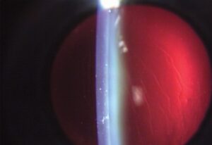
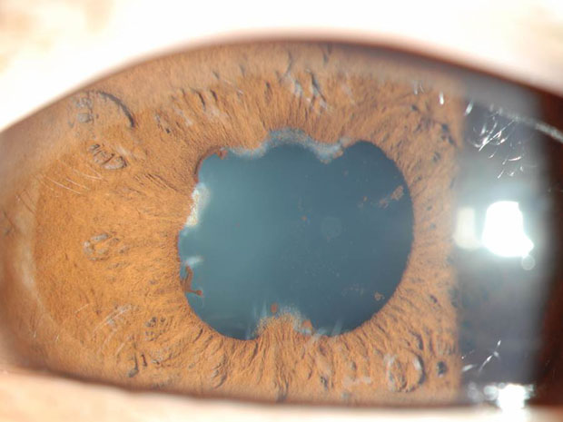
Chronic inflammation causes cataract by itself and by the effect of corticosteroids, and the lens opacity is often posterior subcapsular.
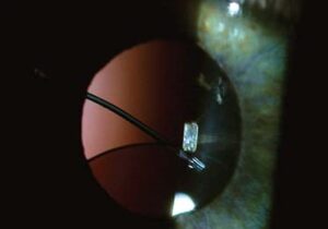
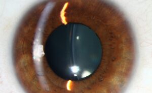
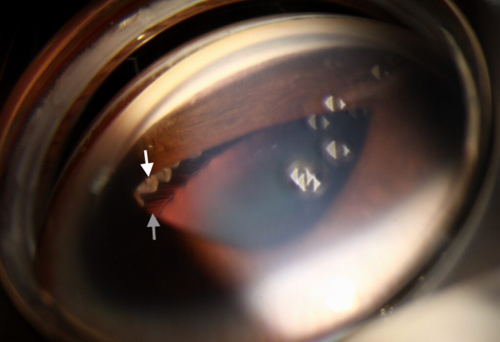
The anterior segment may show posterior synechiae, pupillary membranes, iris atrophy, and band keratopathy (see posterior synechiae).
The blood-aqueous barrier is damaged, so surgery provokes a stronger inflammatory response and fibrin formation.
Macular edema, epiretinal membrane, and glaucoma are common coexisting problems (see uveitic macular edema and uveitic glaucoma).
The result depends as much on the control of inflammation as on the surgery.
Timing
Most surgeons wait until the eye has been quiet for at least three months, with no cells in the anterior chamber. They also want a stable regimen.
The exact interval varies by diagnosis, and some conditions, such as Fuchs heterochromic iridocyclitis, can be operated on in a relatively quiet eye with excellent results.
Surgery during active inflammation is reserved for cases in which the cataract obscures the view of the fundus. It then prevents treatment of a sight-threatening posterior segment disease.
Fundus Explorer Pro
Photograph the retinal findings described here with the phone already in your pocket — 22 D optics and built-in illumination in one handheld unit.
From Choroida — the team behind this sitePerioperative Anti-Inflammatory Cover
- Oral prednisone, often 0.5 to 1 milligram per kilogram daily for several days before and after surgery in patients at high risk
- Intravenous methylprednisolone at the time of surgery in selected severe cases
- Periocular or intravitreal triamcinolone or a dexamethasone implant at the end of the operation
- Topical steroids and nonsteroidal drops, started before surgery and continued for weeks, with a slow taper
- Optimization of immunosuppressive therapy with the uveitis specialist
Patients on systemic steroids need the same attention to pressure, glucose, and bone health as any long-term user.
Surgical Adjustments
- Synechiolysis and pupil enlargement, by viscoelastic, stretching, or a ring or hooks, since miosis is common (see floppy iris syndrome)
- A large capsulorhexis, which helps to avoid capsular phimosis and contraction, though the capsule is fragile
- Careful removal of cortex and polishing of the posterior capsule, to reduce opacification
- Gentle handling of the iris, to avoid bleeding and a fibrinous reaction
- Consideration of posterior capsulotomy and anterior vitrectomy in children and in severe cases
- Lens choice, with a hydrophobic acrylic IOL as the first option in adults, because it has a low rate of cell deposition and capsule opacification
In juvenile idiopathic arthritis uveitis, practice varies.
Some surgeons avoid intraocular lens implantation or choose it selectively, because of the risk of inflammation and complications (see juvenile idiopathic arthritis uveitis).
Outcomes and Complications of Cataract Surgery in Uveitis
Visual outcomes are good in most patients, but they depend on the underlying diagnosis and on the state of the macula and the optic nerve.
- Macular edema is the commonest limiting factor, and its risk is greater in active uveitis
- Posterior capsule opacification is frequent and responds to laser capsulotomy (see YAG laser capsulotomy)
- Postoperative inflammation, fibrin, and synechiae occur more often
- Glaucoma and hypotony can complicate the course
A study using OCT found that macular edema after cataract surgery was more frequent in patients with uveitis than in controls, which supports aggressive prophylaxis.
Follow-Up for Cataract Surgery in Uveitis
Review the patient early, within a day, a week, and then regularly for months, with OCT of the macula and pressure checks.
Taper steroids slowly, and watch for rebound inflammation.
Explain to the patient that visual recovery may be slower and that further treatment may be needed.
Specific Diagnoses
In HLA-B27 anterior uveitis and sarcoidosis, the results are generally good when inflammation is quiet, and perioperative steroid cover is advisable.
In Behçet disease and in severe posterior uveitis, the retina may limit the visual result. So evaluate the fundus and the macula by OCT before promising improvement (see Behçet uveitis).
For herpetic uveitis, antiviral prophylaxis may be needed, and in toxoplasmosis the scar location determines the outlook (see ocular toxoplasmosis).
Counselling
Explain that the surgery is more likely to need extra procedures, that inflammation may flare, and that the final vision depends on the whole eye.
Agree on a plan with the uveitis team for medication before and after surgery. Make sure the patient knows to report pain or redness without delay.
Document the date of the last active inflammation, the drugs used, and the plan for steroid taper in the chart, so that everyone involved in the care knows the timetable.
For the latest studies, see the PubMed results for cataract surgery in uveitis.


Document what you see
Two smartphone imaging tools built for everyday clinic use — one for the slit lamp, one for the fundus.
From Choroida — the team behind this siteReferences
- Bélair ML, Kim SJ, Thorne JE, et al. Incidence of cystoid macular edema after cataract surgery in patients with and without uveitis using optical coherence tomography. Am J Ophthalmol. 2009;148:128-135.
- Okhravi N, Lightman SL, Towler HM. Assessment of visual outcome after cataract surgery in patients with uveitis. Ophthalmology. 1999;106:710-722.
- Foster CS, Vitale AT. Diagnosis and Treatment of Uveitis. 2nd ed. Jaypee; 2013.
- American Academy of Ophthalmology. Basic and Clinical Science Course, Section 9: Uveitis and Ocular Inflammation. AAO; current edition.

Writes and reviews the clinical articles on Educate. Every post is checked against peer-reviewed sources before it goes live.