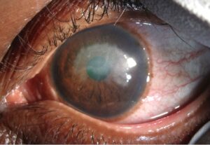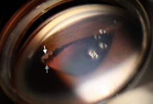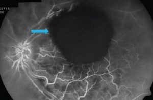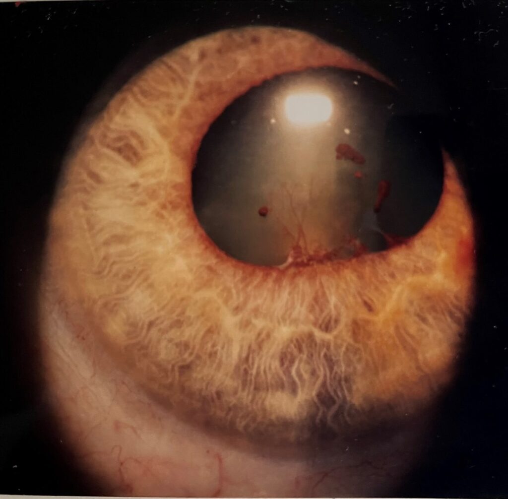
Posterior synechiae are adhesions between the posterior surface of the iris and the anterior lens capsule, most often left behind by anterior uveitis. Fibrin and inflammatory cells in the aqueous organize into fixed bands, and a mature adhesion is usually permanent. Extensive posterior synechiae distort the pupil and can block aqueous flow, leading to secondary angle-closure glaucoma.
They matter because, unlike cells and flare, synechiae do not fade when inflammation settles. A fresh adhesion can sometimes be broken early, but a mature one stays. The shape of the pupil therefore records how much damage past flares have left behind.
What Are Synechiae in the Eye?
A synechia (plural synechiae) is an adhesion between the iris and a neighboring structure in the eye. Two types account for nearly all cases: posterior synechiae, where the iris sticks to the lens, and anterior synechiae, where it sticks to the cornea or the drainage angle. Both follow inflammation, trauma, or surgery, but they cause different problems.
Difference Between Anterior and Posterior Synechiae
Anterior synechiae pull the iris forward to the cornea or trabecular meshwork, while posterior synechiae tether it backward to the lens capsule. Anterior adhesions that close the angle are called peripheral anterior synechiae (PAS) and damage the outflow pathway directly. Posterior adhesions distort the pupil and can trap aqueous behind the iris.
- Posterior synechiae: iris to anterior lens capsule, typically after anterior uveitis, seen as an irregular pupil that dilates poorly.
- Anterior synechiae: iris to cornea or angle, seen after angle closure, flat chamber, trauma, or neovascular disease, and best confirmed by gonioscopy.
All-fit Slit-Lamp Adapter
Record and share exactly what you see at the slit lamp. One adapter fits any slit lamp or surgical microscope — and any smartphone.
From Choroida — the team behind this siteWhy Is the Iris Stuck to the Cornea?
An iris stuck to the cornea is an anterior synechia. It usually follows a flat or shallow anterior chamber, corneal perforation, penetrating trauma, or chronic angle closure. Iridocorneal endothelial syndrome and neovascular membranes can also drag the iris forward, and the resulting angle closure raises intraocular pressure.
Synechiae Formation: How Posterior Synechiae Develop
During active anterior uveitis, breakdown of the blood-aqueous barrier lets fibrin, inflammatory cells, and protein leak into the aqueous. The iris rests against the anterior lens capsule at the pupil margin, so that exudate can organize into a fibrous bond within days if the pupil stays small and immobile.
Chronic or recurrent disease produces the most extensive adhesions, because each flare adds new bands to the old ones. Common causes include HLA-B27 associated acute anterior uveitis, sarcoid uveitis, juvenile idiopathic arthritis-associated uveitis, Behçet disease, and herpetic uveitis.
Because adhesion is time dependent, cycloplegia starts early in a flare rather than waiting for visible synechiae. Keeping the pupil mobile is the most effective way to prevent them.
Posterior Synechiae at the Slit Lamp
At the slit lamp, posterior synechiae appear as pigmented or fibrous strands tethering the pupil margin to the lens capsule. Compare the pupil before and after dilation: adherent segments stay fixed while free segments open, producing a festooned, scalloped pupil. Look for cells, flare, and keratic precipitates to judge current activity.
- Irregular, festooned pupil margin instead of a round contour
- Poor or asymmetric dilation, since adherent iris cannot move
- Pigment on the anterior lens capsule where an adhesion once sat, sometimes remaining after the band has broken
- Adhesion around the entire pupil margin in advanced disease
A festooned pupil in a patient with a remote red eye should prompt questions about prior uveitis, because the synechiae may be the only remaining sign of an episode the patient never took seriously. Record pupil shape and the clock hours of adhesion at each visit so progression is easy to see.
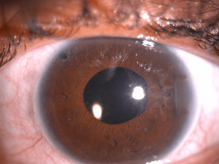
Total Posterior Synechiae, Seclusio Pupillae, and Iris Bombe
Total posterior synechiae, also called seclusio pupillae, means the iris is adherent to the lens for the full 360 degrees of the pupil margin. Aqueous made by the ciliary body then cannot pass through the pupil into the anterior chamber. Pressure builds behind the iris and pushes it forward.
That forward bowing is iris bombe. The peripheral iris is pressed against the trabecular meshwork, the angle closes, and intraocular pressure can rise sharply. The iris looks convex, and the anterior chamber is deep centrally but shallow peripherally.
Iris Bombe vs Posterior Synechiae
Posterior synechiae are the cause, and iris bombe is the consequence. A few synechiae with a free pupil margin do not produce bombe, because aqueous still escapes through the gaps. Bombe needs complete or nearly complete seclusion. Its mechanism differs from primary angle closure, since inflammation rather than crowded anatomy drives it, so treating the inflammation matters as much as lowering pressure.
Differential Diagnosis of an Irregular Pupil
- Traumatic sphincter tear: history of blunt trauma, with a peaked or scalloped margin from focal tears rather than adhesion
- Iris coloboma: congenital, typically inferonasal, present from birth
- Prior intraocular surgery with peripheral iridectomy or iridotomy
- Neovascular glaucoma with ectropion uveae: fibrovascular membrane pulls the pupil margin anteriorly instead of adhering it posteriorly
- Adie tonic pupil: sluggish, segmental reaction with light-near dissociation and no history of intraocular inflammation
Management and Prevention
Cycloplegic agents such as cyclopentolate, homatropine, or atropine, chosen by severity, keep the pupil mobile and prevent synechiae from forming or extending. They also relieve ciliary spasm and pain, which is why they are standard in anterior uveitis alongside topical corticosteroids.
A fresh synechia can sometimes be broken with intensive cycloplegia and topical corticosteroids in the first days, before it organizes. Established adhesions rarely release and may need surgical synechiolysis, often at the time of cataract surgery. Prompt treatment at the start of a flare gives the best chance of avoiding permanent scarring.
Iris bombe with impending or established angle closure needs urgent laser peripheral iridotomy, or surgical iridectomy when laser is not feasible, to create a new route for aqueous. Intensive anti-inflammatory therapy and aqueous suppressants lower pressure meanwhile. Avoid miotics such as pilocarpine, which worsen adhesion and inflammation.
Over the long term, control the underlying uveitis, adding steroid-sparing therapy when flares recur, and check intraocular pressure at every visit in any eye with synechiae. Even incomplete synechiae can extend with later flares, and each new adhesion raises the cumulative risk of angle closure.


Document what you see
Two smartphone imaging tools built for everyday clinic use — one for the slit lamp, one for the fundus.
From Choroida — the team behind this siteReferences
- Jones NP. Glaucoma in uveitis. Eye.
- Moorthy RS, Mermoud A, Baerveldt G, et al. Glaucoma associated with uveitis. Survey of Ophthalmology.
- American Academy of Ophthalmology. Basic and Clinical Science Course, Section 9: Intraocular Inflammation and Uveitis.
- Sallam A, Sheth HG, Habot-Wilner Z, Lightman S. Outcome of raised intraocular pressure in uveitic eyes with and without a corticosteroid-induced hypertensive response. American Journal of Ophthalmology.
Test yourself
A few questions straight from this article.
-
Posterior synechiae are adhesions between which two structures?
Inflammatory fibrin and cellular debris organise where the back of the iris lies against the front of the lens, fixing the two together. -
Which process allows posterior synechiae to form during active anterior uveitis?
Breakdown of the blood-aqueous barrier floods the anterior chamber with fibrin, cells and protein, which can organise into fibrous adhesion within days if the pupil stays constricted and still. -
What term describes posterior synechiae extending around the entire pupil margin?
Seclusio pupillae is complete circumferential adhesion, which prevents any aqueous passing from the posterior to the anterior chamber through the pupil. -
Which sign can persist on the lens after a posterior synechia has been broken?
Pigment left at the former site of adhesion marks where iris and capsule were joined, and remains visible even after the synechia itself is released. -
What sequence of events produces iris bombe in an eye with seclusio pupillae?
With the pupillary route blocked, pressure builds in the posterior chamber and the iris bows into a convex shape, pressing its periphery against the trabecular meshwork and closing the angle. -
What is the urgent treatment for acute iris bombe with angle closure in a uveitic eye?
An iridotomy creates an alternative route for aqueous and relieves the pressure difference between the chambers; the inflammation driving it must be treated at the same time. -
Besides relieving ciliary spasm and pain, why is cycloplegia standard therapy in active anterior uveitis?
Because adhesions organise in a still, constricted pupil within days, cycloplegics are started early and proactively rather than once adhesions are already visible. -
Which approach can still break a posterior synechia that formed only days earlier?
A fresh adhesion can sometimes be broken before it fully organises, which is why treatment at the very start of a flare offers the best chance of avoiding permanent pupillary scarring. -
Which cause of an irregular pupil pulls the pupil margin anteriorly rather than adhering it posteriorly?
In ectropion uveae a fibrovascular membrane drags the pupil margin forward, which is the opposite geometry to a posterior synechia. -
Which feature distinguishes Adie's tonic pupil from posterior synechiae as a cause of pupil irregularity?
The Adie pupil is irregular and sluggish with characteristic light-near dissociation, but there is no preceding intraocular inflammation to have formed adhesions.

Writes and reviews the clinical articles on Educate. Every post is checked against peer-reviewed sources before it goes live.