
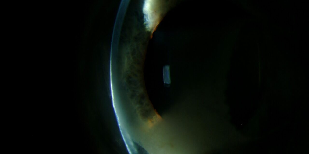
Radiation keratopathy has a particular characteristic that makes it easy to overlook clinically: the corneal complications of radiotherapy often do not appear until months to years after treatment has concluded, meaning that by the time symptoms develop, the connection to a remote course of radiation treatment for an orbital or periocular tumor may not be the first explanation that comes to mind.
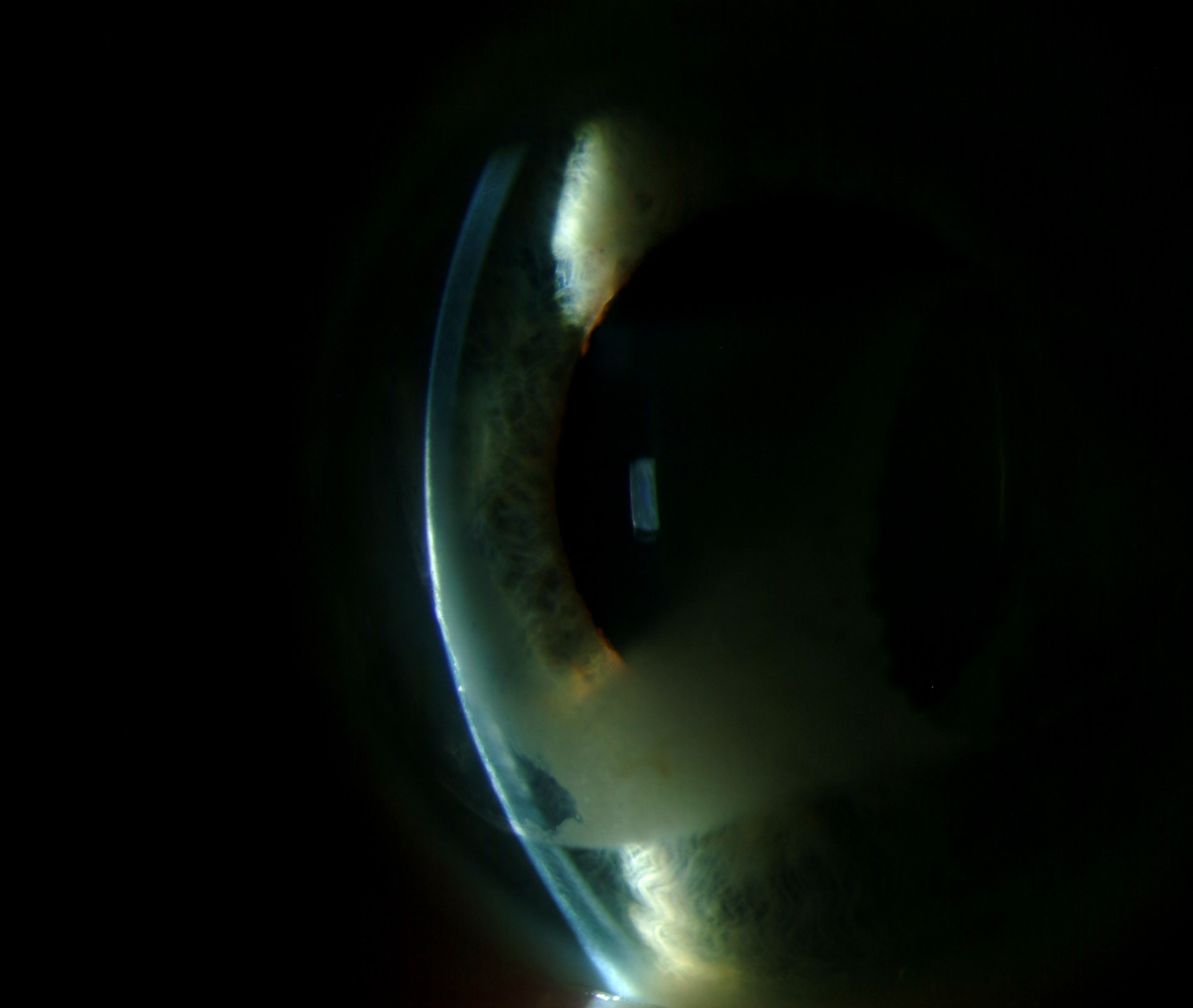
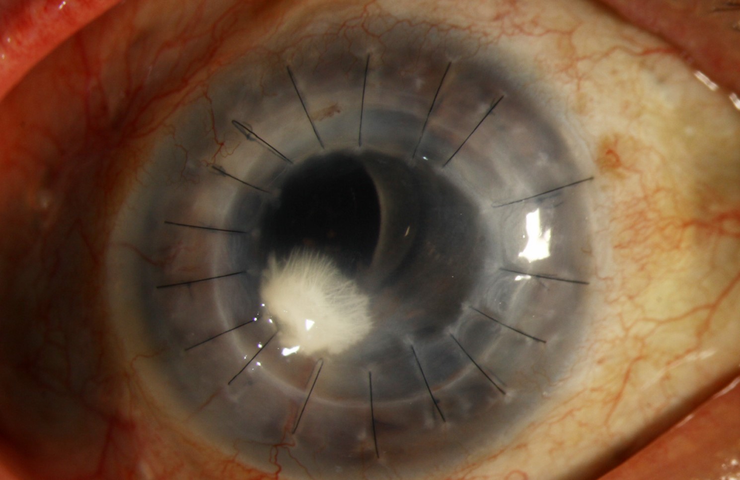
A Delayed, Dose-Dependent Injury
Radiotherapy directed at orbital, periocular, or adjacent structures, used for a range of tumors including orbital lymphoma, lacrimal gland tumors, and periocular skin cancers, can produce corneal and ocular surface injury as a delayed complication, with the severity generally related to the total radiation dose received by ocular structures and, importantly, often not becoming clinically apparent until a considerable interval after treatment has been completed.
Mechanisms of Injury
- Damage to the lacrimal gland reduces aqueous tear production, producing a radiation-induced dry eye that can range from mild to severe depending on the dose received by lacrimal tissue
- Limbal stem cell damage, when radiation dose to the limbal region is significant, can produce limbal stem cell deficiency with its associated corneal surface consequences (see limbal stem cell deficiency classification for the broader discussion of this consequence when it occurs from any cause, including radiation)
- Direct corneal stromal and endothelial effects can occur with higher radiation doses, contributing to corneal edema or scarring in more severely affected cases
- Meibomian gland damage contributes an evaporative dry eye component alongside any aqueous deficiency from lacrimal gland injury
All-fit Slit-Lamp Adapter
Record and share exactly what you see at the slit lamp. One adapter fits any slit lamp or surgical microscope — and any smartphone.
From Choroida — the team behind this siteClinical Features
- Dry eye symptoms, often progressive, developing over the months to years following radiotherapy, sometimes not recognized as radiation-related given the delayed onset relative to the original treatment
- Punctate keratopathy, and in more severe cases, persistent epithelial defects or corneal ulceration, reflecting the cumulative impact of tear deficiency and any associated limbal stem cell compromise
- In cases with significant limbal involvement, conjunctivalization and vascularization of the corneal surface can develop, following the same pattern seen with limbal stem cell deficiency from other causes
- Eyelid changes, including lash loss (madarosis) and lid margin keratinization, can accompany the ocular surface findings, reflecting the broader periocular tissue effects of radiation exposure
Why the Delayed Onset Matters Clinically
Because radiation keratopathy can present a considerable time after the causative treatment, a relevant radiation history should be specifically sought in any patient presenting with unexplained, progressive dry eye or ocular surface disease, particularly one with a history of orbital, periocular, or nasal/sinus tumor treatment, even when that treatment occurred years earlier and might not otherwise be volunteered as relevant by the patient.
Diagnostic Evaluation
- A specific history of any prior orbital, periocular, or adjacent-structure radiotherapy, including approximate dose and treatment field when this information is available, given the delayed-onset nature of this complication
- Comprehensive dry eye and ocular surface evaluation, including tear film assessment and ocular surface staining
- Assessment for limbal involvement and any resulting conjunctivalization, particularly in patients who received higher radiation doses or had treatment fields closer to the limbus
Management
Ocular Surface Support
Management follows similar principles to severe dry eye and ocular surface disease from other causes, including preservative-free lubrication, punctal occlusion, and topical anti-inflammatory treatment as needed, tailored to the severity of findings in a given patient.
Managing Limbal Stem Cell Compromise
When significant limbal stem cell deficiency has resulted from radiation exposure, management follows the same staged approach used for this condition from other causes, potentially including limbal stem cell transplantation for more severe, visually significant cases (see limbal stem cell transplantation techniques for the relevant surgical management framework).
Ongoing Surveillance
Given the potential for slow, progressive worsening over an extended period following radiation exposure, ongoing periodic monitoring, rather than a single evaluation and discharge, is appropriate for patients with a significant history of orbital or periocular radiotherapy, allowing earlier detection and management of progressive changes over time.
Prognosis
Radiation keratopathy severity and prognosis vary considerably depending on the radiation dose received by ocular structures and the specific structures affected, with milder cases responding well to standard ocular surface support measures and more severe cases, particularly those with significant limbal stem cell loss, requiring more extensive reconstructive management.
Awareness of this condition’s characteristic delayed presentation supports earlier recognition in patients presenting with otherwise unexplained ocular surface disease, allowing more prompt initiation of appropriate supportive treatment before significant cumulative corneal damage develops.


Document what you see
Two smartphone imaging tools built for everyday clinic use — one for the slit lamp, one for the fundus.
From Choroida — the team behind this siteReferences
- Kim JH, Chung H, Choi MY, et al. Ocular complications of radiotherapy for periocular tumors. Surv Ophthalmol. 2014;59:363-375.
- Parsons JT, Bova FJ, Fitzgerald CR, Mendenhall WM, Million RR. Radiation optic neuropathy after megavoltage external-beam irradiation: analysis of time-dose factors. Int J Radiat Oncol Biol Phys. 1994;30:755-763.
- Merriam GR Jr, Szechter A, Focht EF. The effects of ionizing radiations on the eye. Front Radiat Ther Oncol. 1972;6:346-385.
- Brady LW, Shields J, Augsburger J, Markoe AM. Complications from radiation therapy to the eye. Front Radiat Ther Oncol. 1989;23:238-250.
Test yourself
A few questions straight from this article.
-
When do the corneal complications of radiation keratopathy typically become apparent?
The delayed onset is the defining feature: corneal injury often surfaces months to years after radiotherapy ends, so the link to the original treatment is easily missed. -
What chiefly determines the severity of radiation keratopathy?
Severity is generally related to the total dose delivered to ocular structures, which is why dose and treatment field are worth establishing when the history is taken. -
Radiotherapy for which tumours characteristically leads to radiation keratopathy?
Radiotherapy aimed at orbital, periocular or adjacent structures for tumours such as orbital lymphoma, lacrimal gland tumours and periocular skin cancers can injure the cornea and ocular surface. -
Which radiation injury produces the aqueous tear deficiency in radiation keratopathy?
Radiation damage to the lacrimal gland reduces aqueous tear production, giving a radiation-induced dry eye whose severity tracks the dose that lacrimal tissue received. -
A significant radiation dose to the limbal region can result in which condition?
Where the limbal dose is significant, limbal stem cell damage can produce limbal stem cell deficiency with its usual corneal surface consequences. -
What corneal surface change develops when radiation keratopathy involves the limbus significantly?
With significant limbal involvement the cornea becomes conjunctivalized and vascularized, following the same pattern as limbal stem cell deficiency from other causes. -
Which eyelid changes can accompany the ocular surface findings of radiation keratopathy?
Madarosis and lid margin keratinization reflect the broader effect of radiation on periocular tissues and often accompany the surface disease. -
Which history should be sought specifically in a patient with unexplained, progressive ocular surface disease?
Because presentation is so delayed, a radiation history for orbital, periocular or nasal and sinus tumours should be asked for directly, since patients may not volunteer treatment from years ago. -
Why is ongoing periodic review preferred to a single assessment after significant periocular radiotherapy?
Slow, progressive worsening can continue long after exposure, so periodic monitoring rather than one evaluation and discharge allows earlier detection of change. -
How is severe, visually significant radiation-induced limbal stem cell deficiency managed?
Radiation-induced limbal stem cell deficiency follows the same staged approach used for other causes, which can extend to limbal stem cell transplantation in severe, visually significant cases.
