
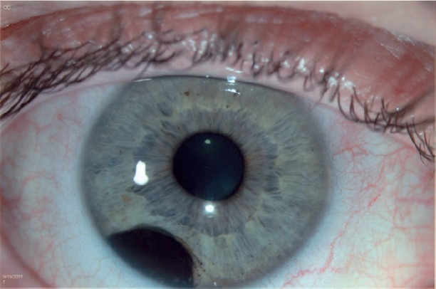
Iris melanoma is the least common but, in an important practical sense, the most visible of the uveal melanomas, arising in the anterior segment where it can be directly seen and photographed at the slit lamp rather than discovered incidentally on a dilated fundus exam the way choroidal melanoma, discussed in its own dedicated article on this site, often is.
This visibility is a genuine clinical advantage, because it allows for the kind of careful serial documentation and growth monitoring that is harder to achieve for a tumor tucked away in the posterior segment.
General Prognosis Relative to Other Uveal Melanoma
Iris melanoma carries a more favorable overall prognosis than choroidal or ciliary body melanoma, related to a combination of factors: it tends to be diagnosed earlier given its visibility, tends to be smaller at diagnosis, and appears to have a somewhat lower intrinsic rate of metastatic spread even accounting for its earlier detection.
This does not mean iris melanoma is without risk — a subset of cases do behave more aggressively and metastasize — but the overall more favorable natural history is a genuine and clinically relevant distinction from its posterior segment counterparts.
Clinical Presentation
Patients or their eye care provider typically notice a pigmented iris lesion, sometimes present for years before any change is documented, and concern arises specifically when the lesion demonstrates growth, develops new features (increased vascularity, irregular pupil, secondary glaucoma), or when a lesion is large or atypical enough at first presentation to raise suspicion without needing to wait for documented change over time.
All-fit Slit-Lamp Adapter
Record and share exactly what you see at the slit lamp. One adapter fits any slit lamp or surgical microscope — and any smartphone.
From Choroida — the team behind this siteExam Findings Suggesting Malignancy
- Documented growth on serial photography — as with choroidal melanoma, this is the single most decisive feature separating an active melanoma from a stable iris nevus
- Prominent, feeder or intrinsic tumor vascularity, visible as abnormal blood vessels on or leading into the lesion
- Secondary glaucoma from tumor-related angle involvement or pigment dispersion, discussed further in this site’s coverage of pigment dispersion and pigmentary glaucoma for the general mechanism, though here driven by the tumor itself rather than a primary pigment dispersion process
- Ectropion uveae (posterior iris pigment epithelium pulled anteriorly onto the iris surface) adjacent to the lesion
- Pupil distortion or irregularity from local tumor infiltration of the iris sphincter or stroma
- A lesion size exceeding roughly 3 mm in diameter or 1 mm in thickness — larger lesions carry a meaningfully higher pretest probability of being melanoma rather than a benign nevus, similar in spirit to the size-based risk stratification used for choroidal lesions
Differential Diagnosis
- Iris nevus — stable, without documented growth or the concerning features listed above; the great majority of pigmented iris lesions encountered in practice are benign nevi rather than melanoma, and most require only periodic monitoring rather than active intervention
- Iris cyst — generally translucent or cystic in appearance on slit-lamp exam, often confirmed with ultrasound biomicroscopy, and discussed in its own dedicated article on this site
- Iris pigment epithelial hyperplasia or hamartoma — benign pigmented lesions with their own characteristic, generally stable appearance, discussed in this site’s coverage of iris hamartomas
- Foreign body or metallic deposition (siderosis) — a relevant trauma history and a different, less discretely mass-like appearance generally distinguish these from a true iris tumor
- Iris metastasis — typically from a known primary malignancy elsewhere, sometimes multifocal, and an important consideration in a patient with a relevant oncologic history presenting with a new iris lesion
Diagnostic Evaluation
Careful slit-lamp examination with photographic documentation establishes a baseline, and periodic re-examination with repeat photography is the primary tool for detecting the growth that most reliably distinguishes melanoma from a stable nevus.
Anterior segment ultrasound biomicroscopy characterizes lesion thickness and any extension into the ciliary body or angle not fully visible on slit-lamp exam alone, and is especially useful for larger or more posteriorly extending lesions where the full extent may not be directly visualized through the pupil.
Management
Small, stable, asymptomatic lesions without concerning features are observed with periodic monitoring and photographic documentation, similar in principle to the observation approach used for a suspicious but not yet growing choroidal lesion.
Documented growth or a lesion with sufficiently concerning features at presentation warrants active treatment: local resection (iridectomy or iridocyclectomy for lesions also involving the ciliary body) is more feasible for anterior segment tumors than for posterior uveal melanoma, given the accessible location, and plaque brachytherapy is also used for appropriately selected lesions, following similar principles to its use in choroidal melanoma discussed elsewhere on this site.
Enucleation is reserved for extensive tumors not amenable to eye-sparing treatment, or those causing uncontrolled secondary glaucoma with no reasonable visual potential remaining.
As with choroidal melanoma, systemic surveillance for metastatic disease continues after local treatment, tailored to the individual tumor’s size and risk features, though the overall lower metastatic potential of iris melanoma compared with its posterior counterparts generally translates into a less intensive, though still important, long-term surveillance approach.
Communicating this comparatively favorable prognosis clearly, while still emphasizing the genuine need for ongoing follow-up, helps patients understand both why iris melanoma is treated seriously and why the outlook here differs meaningfully from that of choroidal or ciliary body melanoma.


Document what you see
Two smartphone imaging tools built for everyday clinic use — one for the slit lamp, one for the fundus.
From Choroida — the team behind this siteReferences
- Shields CL, Kaliki S, Hutchinson A, et al. Iris nevus growth into melanoma: analysis of 1611 consecutive eyes. Ophthalmology.
- Shields JA, Shields CL. Iris melanoma: risk factors for metastasis. Community Eye Health Journal.
- American Academy of Ophthalmology. Basic and Clinical Science Course, Section 4: Ophthalmic Pathology and Intraocular Tumors.
Test yourself
A few questions straight from this article.
-
Iris melanoma differs from the other uveal melanomas chiefly in which respect?
Iris melanoma is the least common uveal melanoma, but because it sits in the anterior segment it can be seen and photographed directly at the slit lamp rather than found incidentally on fundus examination. -
How does the overall prognosis of iris melanoma compare with choroidal and ciliary body melanoma?
Iris melanoma is usually diagnosed earlier and smaller, and appears to have a somewhat lower intrinsic rate of metastatic spread, giving it a more favorable natural history than its posterior counterparts. -
Which single feature most reliably separates an active iris melanoma from a stable iris nevus?
As with choroidal melanoma, documented growth is the single most decisive feature distinguishing melanoma from a stable nevus, which is why baseline and repeat slit-lamp photography matter so much. -
Above roughly what size does a pigmented iris lesion carry a higher probability of melanoma?
Lesions exceeding roughly 3 mm in diameter or 1 mm in thickness carry a meaningfully higher pretest probability of melanoma, echoing the size-based risk stratification used for choroidal lesions. -
Which slit-lamp finding on a pigmented iris lesion points to melanoma rather than a benign nevus?
Abnormal vessels on or leading into the lesion are listed among the features suggesting malignancy, alongside secondary glaucoma, ectropion uveae and pupil distortion. -
What is true of most pigmented iris lesions encountered in routine ophthalmic practice?
The great majority of pigmented iris lesions are benign nevi that remain stable and require nothing more than periodic monitoring rather than active intervention. -
Which investigation best measures iris lesion thickness and extension into the angle or ciliary body?
Ultrasound biomicroscopy characterises lesion thickness and any ciliary body or angle extension that cannot be fully seen through the pupil, and also helps confirm a suspected iris cyst. -
How is a small, stable, asymptomatic iris lesion without concerning features managed?
Small, stable, asymptomatic lesions are observed with periodic monitoring and photography, the same principle applied to a suspicious but non-growing choroidal lesion. -
Which resection is used for an iris melanoma that also involves the ciliary body?
Local resection is more feasible for anterior segment tumours; iridectomy suffices for iris-confined disease, while iridocyclectomy is used when the ciliary body is also involved. -
For which iris melanomas is enucleation reserved?
Enucleation is kept for tumours too extensive for eye-sparing treatment, or those causing uncontrolled secondary glaucoma in an eye with no reasonable visual potential.

Writes and reviews the clinical articles on Educate. Every post is checked against peer-reviewed sources before it goes live.


