
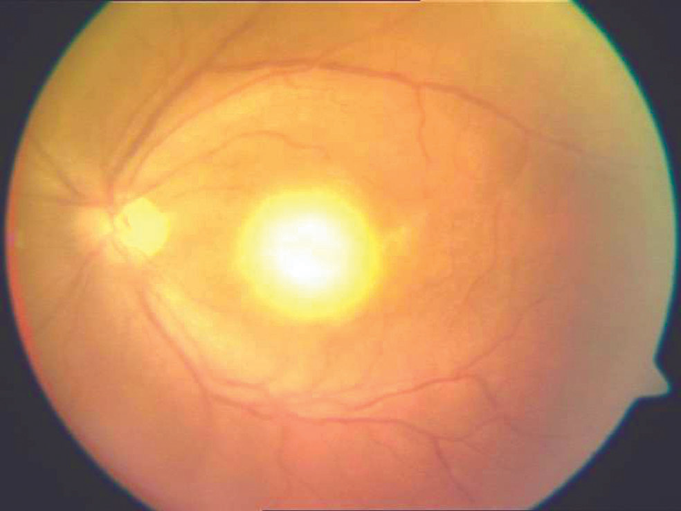
Ocular toxoplasmosis is retinochoroiditis caused by the protozoan parasite Toxoplasma gondii. It is the most common cause of infectious posterior uveitis worldwide.
In a patient with the right fundus appearance, it is often recognisable before the workup even begins.
Infection is acquired in one of two ways. Congenital infection is transmitted transplacentally when a mother is infected during pregnancy; the risk of transmission and the severity of resulting disease depend on the trimester in which maternal infection occurred, with later infection transmitting more often but generally causing milder disease.
Acquired infection later in life comes from contaminated food, water, or contact with cat faeces, the parasite’s definitive host being the domestic cat. What is now well established, though not always appreciated by trainees, is that most of the ocular disease seen in clinic is not new infection at all but reactivation of an old, previously silent chorioretinal scar.
What Is Ocular Toxoplasmosis?
Toxoplasmosis is caused by tachyzoites invading retinal cells and producing a focal, necrotising retinitis. The choroid becomes secondarily inflamed adjacent to the primary retinal focus, which is why the disease is described as retinochoroiditis rather than simple retinitis.
As the active lesion heals, the retina and RPE are replaced by gliotic tissue, leaving a well-demarcated, pigmented chorioretinal scar.
The clinically important detail is what happens inside that scar afterwards. Bradyzoite cysts persist within the healed tissue indefinitely and can reactivate at the margin of the old scar, producing a new adjacent focus of active retinitis.
This is the basis of the classic “satellite lesion” pattern that experienced clinicians recognise on sight: a fresh, fluffy white area of active retinitis sitting directly beside an old, sharply demarcated pigmented scar. Reactivation almost always occurs at the edge of an existing scar rather than at a new, unrelated site, so the location of a patient’s first lesion is a reasonable predictor of where future recurrences will appear.
Epidemiology
Seroprevalence for Toxoplasma gondii varies widely by region and diet, and only a minority of seropositive individuals ever develop ocular disease. Recurrence, however, is common in those who do, and each episode adds to the cumulative burden of scarring.
Immunocompromised patients, including those with HIV/AIDS, are prone to more atypical, bilateral, and severe disease, and this group deserves a lower threshold for aggressive treatment and closer follow-up.
Fundus Explorer Pro
Photograph the retinal findings described here with the phone already in your pocket — 22 D optics and built-in illumination in one handheld unit.
From Choroida — the team behind this siteClinical Presentation
Patients typically present with blurred vision and floaters reflecting vitreous inflammatory debris. Pain, redness, and photophobia can accompany significant anterior chamber reaction, and a central or paracentral scotoma develops when the lesion involves or threatens the macula or papillomacular bundle.
Congenital disease may present in infancy with strabismus or a white pupillary reflex noticed by a parent, though it is just as often picked up incidentally on a later examination when a scar is found that no one knew was there.

On examination, the active lesion appears as a focal, fluffy white or yellow-white area of retinitis, classically adjacent to a pigmented chorioretinal scar. Overlying vitritis gives the lesion its hazy, indistinct border, which is where the “headlight in fog” description comes from: a bright focal lesion seen through a diffusely hazy vitreous.
Retinal vasculitis with vascular sheathing can be seen near the active focus in more severe cases, and granulomatous keratic precipitates with anterior chamber reaction sometimes accompany the posterior findings. Old, healed lesions, in contrast, are sharply demarcated, atrophic, and pigmented, with no overlying haze at all.
In a patient from an area with reasonable background seroprevalence, an active fluffy lesion sitting next to an old pigmented scar is close to diagnostic on its own, and many cases are treated on this appearance without further testing.
Diagnostic Evaluation
IgG and IgM serology is useful mainly for excluding the diagnosis: ocular toxoplasmosis in a seronegative patient would be very unusual, so a negative IgG argues strongly against it.
A positive IgG, on the other hand, only confirms past exposure and does not prove that the lesion in front of you is active toxoplasmosis, particularly in populations where background seroprevalence is high.
Aqueous or vitreous PCR, together with calculation of the Goldmann-Witmer coefficient, can confirm the diagnosis in atypical presentations or in immunocompromised patients where the clinical picture is less clear. This level of testing is not needed in most typical cases.
OCT is useful for assessing macular involvement and epiretinal membrane formation, and fluorescein angiography typically shows early hypofluorescence of the active lesion with late staining, plus vasculitis where present.
Differential Diagnosis
- Toxocariasis, which usually presents as a solitary granuloma without an adjacent old scar, often in a younger child with a different exposure history
- Acute retinal necrosis, which tends to be more rapidly progressive with confluent peripheral necrosis and marked vasculitis, typically in an immunocompromised or herpesvirus-infected patient
- Cytomegalovirus retinitis, with its haemorrhagic “pizza pie” appearance, seen in significantly immunocompromised patients
- Syphilitic chorioretinitis, which can look almost anything, and should be excluded serologically whenever the picture is atypical
- Sarcoid or tuberculous chorioretinitis, where the systemic context points toward the diagnosis
The presence of an old, adjacent scar is the single most useful feature separating toxoplasmosis from this list, since most of these other entities do not produce that specific satellite configuration.
Management
Not every lesion needs treatment. Small peripheral lesions with mild inflammation and no threat to central vision are often observed, since the disease is frequently self-limiting.
Treatment is indicated when the lesion threatens the macula, optic nerve, or papillomacular bundle, when vitritis is significant enough to reduce vision meaningfully, or in immunocompromised patients, who need more aggressive management given their risk of severe or atypical disease.
Classic triple therapy with pyrimethamine, sulfadiazine, and folinic acid remains a standard regimen, though it requires monitoring for haematological toxicity over the treatment course. Trimethoprim-sulfamethoxazole has become a widely used alternative with comparable efficacy and a more favourable side-effect profile in several comparative studies.
Clindamycin, azithromycin, and atovaquone are used as alternative or adjunctive agents, particularly where sulfa allergy is a concern. Treatment typically runs four to six weeks.
Systemic corticosteroids are often added for sight-threatening lesions, but only once antiparasitic therapy is already underway. Steroids should not be used as monotherapy: suppressing the inflammatory response without covering the underlying infection risks unchecked parasitic proliferation and a worse outcome.
This sequencing, antiparasitic cover first, is the one prescribing point in this disease that is worth getting right every time.
Prognosis
Peripheral lesions away from the macula tend to resolve with a good visual outcome, leaving a scar that carries little functional consequence. Macular or juxtapapillary lesions are a different matter: scarring in these locations causes permanent central or paracentral field loss regardless of how well the acute inflammation is controlled.
Recurrence is common over a patient’s lifetime, and because each episode can add scarring near the fovea, counselling patients about ongoing surveillance is as much a part of management as treating the current episode.
Congenital disease identified and treated early in infancy generally does better in the long run than disease that is allowed to declare itself later.


Document what you see
Two smartphone imaging tools built for everyday clinic use — one for the slit lamp, one for the fundus.
From Choroida — the team behind this siteReferences
- Holland GN. Ocular toxoplasmosis: a global reassessment. American Journal of Ophthalmology. 2003.
- Felix JP, Lira RP, Zacchia RS, et al. Trimethoprim-sulfamethoxazole versus pyrimethamine and sulfadiazine for ocular toxoplasmosis. American Journal of Ophthalmology. 2014.
- Butler NJ, Furtado JM, Winthrop KL, Smith JR. Ocular toxoplasmosis II: clinical features, pathology and management. Clinical and Experimental Ophthalmology. 2013.
- Ocular Toxoplasmosis. EyeWiki, American Academy of Ophthalmology.
- Toxoplasmosis, Ocular. StatPearls, NCBI Bookshelf.
Test yourself
A few questions straight from this article.
-
Ocular toxoplasmosis is the most common cause of which group of eye disorders worldwide?
Retinochoroiditis caused by Toxoplasma gondii is the most common cause of infectious posterior uveitis worldwide. -
What explains recurrence of ocular toxoplasmosis at the edge of an old chorioretinal scar?
Bradyzoite cysts persist indefinitely inside the healed scar and reactivate at its margin, producing a new adjacent focus of active retinitis. -
What creates the 'headlight in fog' appearance in ocular toxoplasmosis?
Overlying vitritis gives the active lesion a hazy, indistinct border, so a bright focal lesion is seen through a diffusely hazy vitreous. -
Which feature typically distinguishes toxocariasis from ocular toxoplasmosis?
Toxocariasis usually presents as a solitary granuloma without an adjacent old scar, and the satellite configuration is the single most useful discriminating feature. -
How should a negative Toxoplasma IgG result be interpreted in suspected ocular toxoplasmosis?
Serology is most useful for exclusion: ocular toxoplasmosis in a seronegative patient would be very unusual, so a negative IgG argues strongly against it. -
What does fluorescein angiography typically show over an active toxoplasmic lesion?
Fluorescein angiography typically shows early hypofluorescence of the active lesion with late staining, plus vasculitis where present. -
Which situation makes treatment of an ocular toxoplasmosis lesion indicated rather than optional?
Treatment is indicated when the lesion threatens the macula, optic nerve or papillomacular bundle, when vitritis meaningfully reduces vision, or in immunocompromised patients. -
When may systemic corticosteroids be added for a sight-threatening toxoplasmic lesion?
Steroids must never be used as monotherapy: suppressing inflammation without covering the infection risks unchecked parasitic proliferation and a worse outcome. -
How long does antiparasitic treatment for ocular toxoplasmosis typically run?
Whether classic triple therapy or trimethoprim-sulfamethoxazole is used, treatment typically runs four to six weeks. -
Why do macular or juxtapapillary toxoplasmic lesions carry a worse visual prognosis?
Scarring in these locations causes permanent central or paracentral field loss regardless of how well the acute inflammation is controlled.

Writes and reviews the clinical articles on Educate. Every post is checked against peer-reviewed sources before it goes live.


