
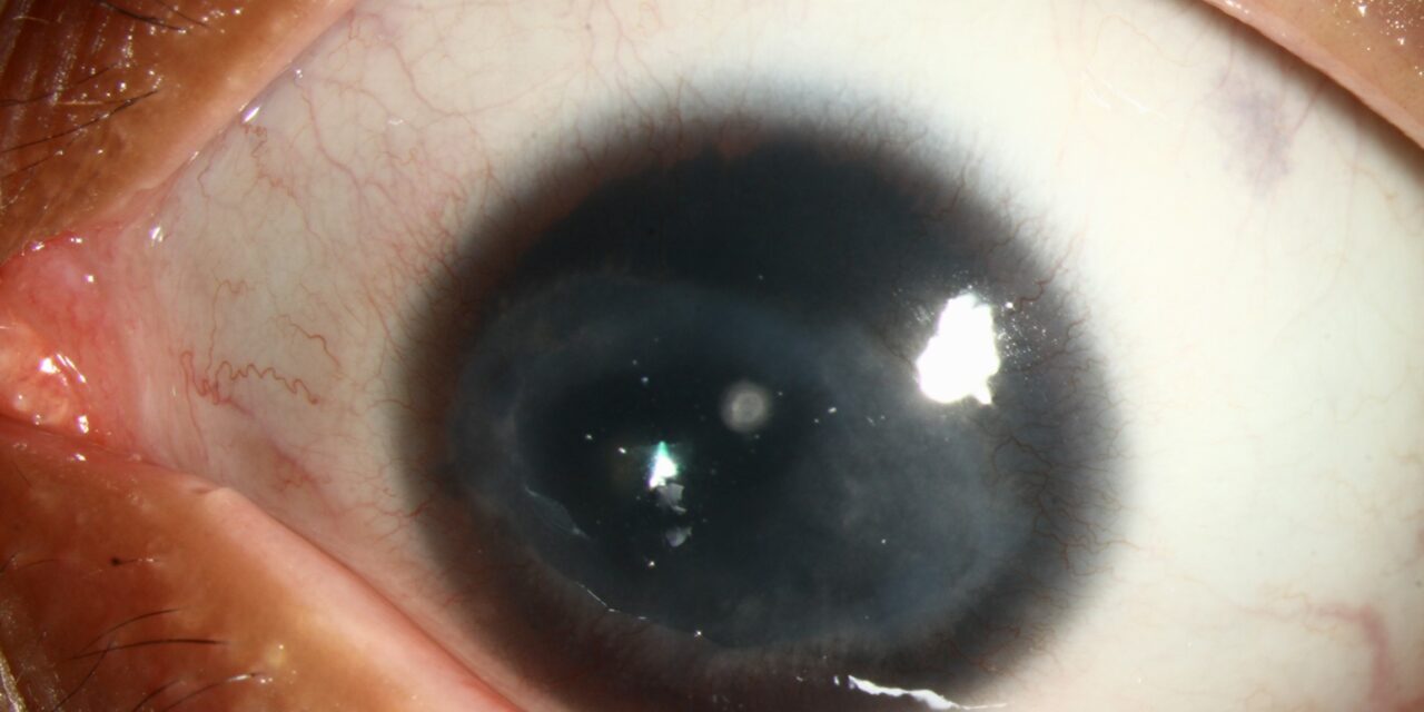
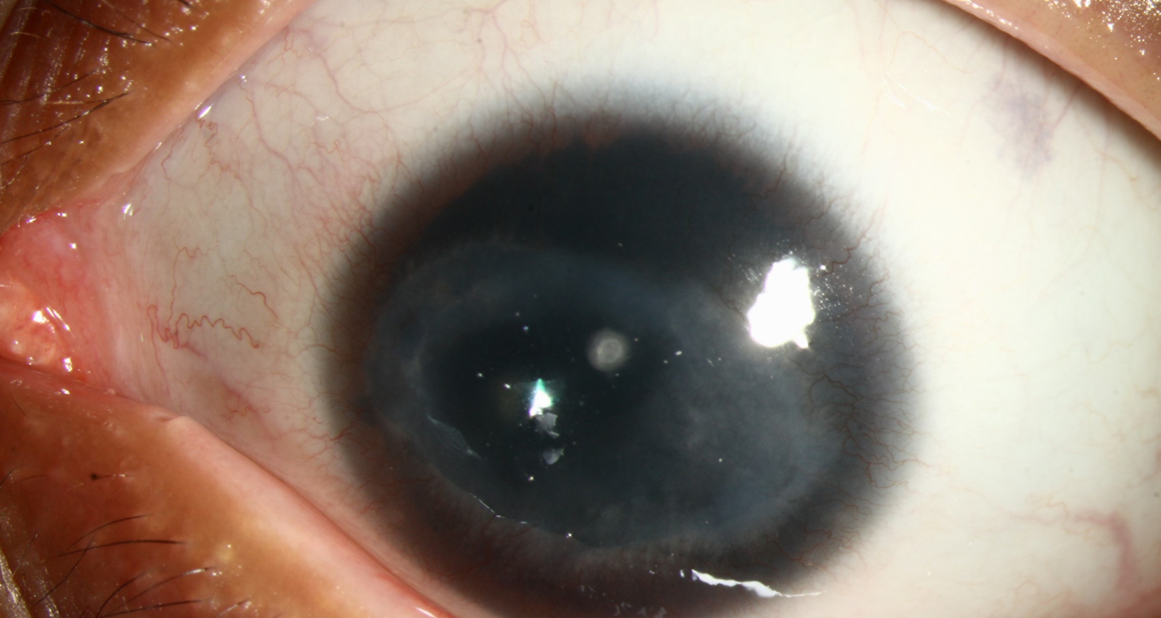
Corneal pannus, the ingrowth of superficial fibrovascular tissue across the normally avascular cornea, is the eye’s response to a cornea that has been chronically deprived of oxygen or chronically inflamed, and because different underlying causes tend to produce vascularization in characteristically different patterns and depths, careful attention to exactly where and how the new vessels are growing can meaningfully narrow the differential diagnosis.
Why the Normal Cornea Has No Blood Vessels
The cornea is normally avascular, a property essential for maintaining its transparency and, by extension, good vision, maintained through a combination of anti-angiogenic factors and the cornea’s relatively low metabolic demand, met by oxygen diffusion from the tear film and aqueous humor rather than requiring its own blood supply.
When this normal avascular state is disrupted, whether by chronic hypoxia, inflammation, or limbal stem cell dysfunction, angiogenic signals overcome the cornea’s normal anti-angiogenic defenses, and new vessels grow in from the limbal vascular arcade, producing pannus, vascularization that, while sometimes a necessary or at least understandable healing response, can itself reduce corneal clarity and vision if extensive.
Superficial Pannus
- Vessels growing within the most superficial corneal layers, typically just beneath the epithelium
- Classically associated with chronic contact lens wear, particularly with extended or overnight wear causing chronic hypoxia (see contact lens overwear syndrome)
- Trachoma, a chronic chlamydial conjunctivitis endemic in some regions of the world, classically produces a superior superficial pannus, historically one of the most recognizable and diagnostically important signs of this condition globally
- Chronic blepharitis and ocular surface inflammation more broadly can also drive superficial vascularization over time
All-fit Slit-Lamp Adapter
Record and share exactly what you see at the slit lamp. One adapter fits any slit lamp or surgical microscope — and any smartphone.
From Choroida — the team behind this siteDeep Stromal Vascularization
- Vessels extending more deeply into the corneal stroma, associated with conditions producing deeper corneal inflammation
- Interstitial keratitis, whether from congenital syphilis, herpes simplex, or other causes, classically produces deep stromal vascularization, sometimes leaving behind non-perfused “ghost vessels” even after the active disease has resolved (see interstitial keratitis)
- Chemical and thermal burns, when severe, can also produce deep vascularization as part of the healing and scarring response
Vascularization From Limbal Stem Cell Deficiency
When the limbal stem cells responsible for maintaining a normal, healthy corneal epithelium are damaged or deficient, the resulting epithelium is often poorly formed and unstable, and this instability, along with loss of the normal limbal barrier function, allows conjunctival tissue, including its blood vessels, to migrate onto and across the corneal surface, a specific vascularization pattern associated with conjunctivalization of the cornea (see limbal stem cell deficiency).
Clinical Evaluation
- Careful assessment of vascularization depth (superficial versus deep stromal), distribution (localized versus circumferential, and which clock hours are involved), and extent, since all of these features contribute diagnostic information
- A directed history covering contact lens wear pattern, prior ocular surface disease or trauma, and any relevant systemic or infectious exposure history
- Assessment of the corneal epithelium for signs of limbal stem cell deficiency when vascularization is associated with an irregular, unstable-appearing epithelial surface
- Documentation with slit-lamp photography, useful both for tracking any progression and for objectively assessing response to treatment over time
Management
Addressing the Underlying Cause
The central principle of managing corneal pannus is treating the underlying driver, whether that means modifying or discontinuing problematic contact lens wear, treating chronic blepharitis or ocular surface inflammation, addressing an underlying infectious cause, or managing limbal stem cell deficiency, since the vascularization itself is a response to an ongoing stimulus rather than an independent disease process.
Anti-Inflammatory and Anti-Angiogenic Treatment
Topical corticosteroids can help reduce active inflammation driving vascularization, and in some cases, targeted anti-angiogenic treatments have been used for problematic, vision-threatening corneal neovascularization, though addressing the underlying cause remains the primary and most durable strategy.
Considerations Before Corneal Transplantation
Established, extensive corneal vascularization is an important consideration when corneal transplantation is being planned for any reason, since vascularized corneas carry a substantially higher risk of graft rejection than avascular corneas, given the more direct access of the host immune system to donor tissue through the established vessels, which is why addressing vascularization, where possible, before proceeding with transplantation improves the likelihood of long-term graft success.
Prognosis
Early, limited pannus, once the underlying cause is identified and addressed, often stabilizes or partially regresses, particularly vascularization related to a reversible cause such as contact lens overwear.
More extensive, longstanding vascularization, particularly when associated with limbal stem cell deficiency or severe chronic inflammatory or infectious disease, is more likely to persist and can meaningfully complicate future corneal surgery, underscoring the value of recognizing and addressing corneal vascularization early, before it becomes extensive and established.


Document what you see
Two smartphone imaging tools built for everyday clinic use — one for the slit lamp, one for the fundus.
From Choroida — the team behind this siteReferences
- Chang JH, Gabison EE, Kato T, Azar DT. Corneal neovascularization. Curr Opin Ophthalmol. 2001;12:242-249.
- Lee P, Wang CC, Adamis AP. Ocular neovascularization: an epidemiologic review. Surv Ophthalmol. 1998;43:245-269.
- Taylor HR. Trachoma: a blinding scourge from the bronze age to the twenty-first century. Melbourne: Haddington Press; 2008.
- Dana MR, Streilein JW. Loss and restoration of immune privilege in eyes with corneal neovascularization. Invest Ophthalmol Vis Sci. 1996;37:2485-2494.
