
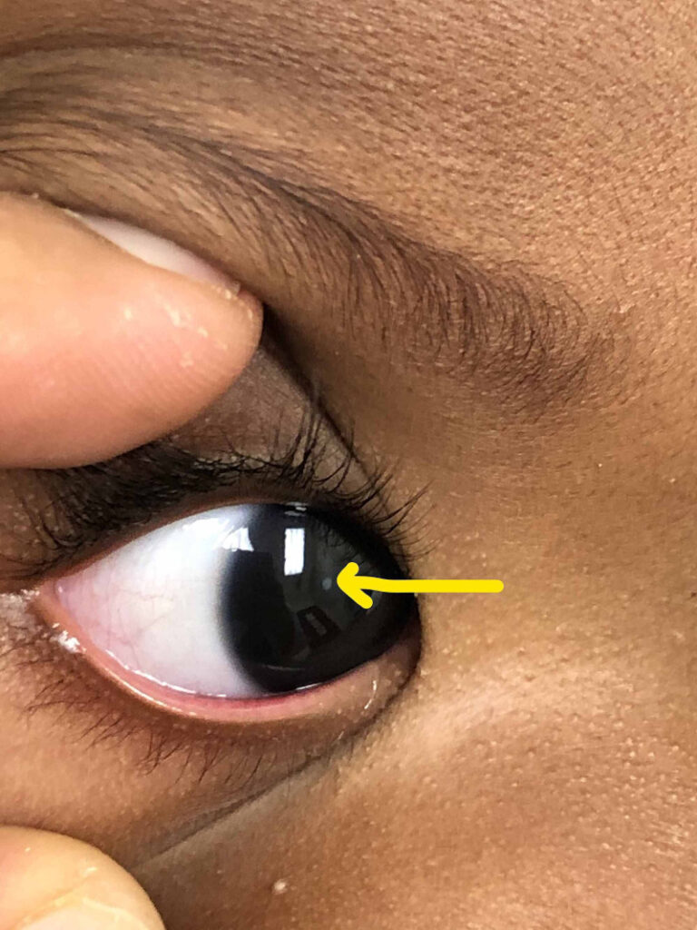
Leukocoria, a white pupillary reflex in place of the normal red reflex, is a physical sign rather than a diagnosis, and its differential spans conditions ranging from an entirely benign cataract to a life-threatening malignancy, which is exactly why any child presenting with this finding needs a systematic, urgent evaluation rather than a casual reassurance.
Retinoblastoma, discussed in its own dedicated article on this site, is the diagnosis every clinician rightly thinks of first when leukocoria is noted, but it is far from the only cause, and organizing the broader differential helps avoid both the danger of missing a malignancy and the opposite error of unnecessary alarm over a benign finding.
A structured approach to this differential, built around a careful history, exam, and targeted imaging, is what actually distinguishes these possibilities reliably rather than relying on the pupil’s appearance alone.
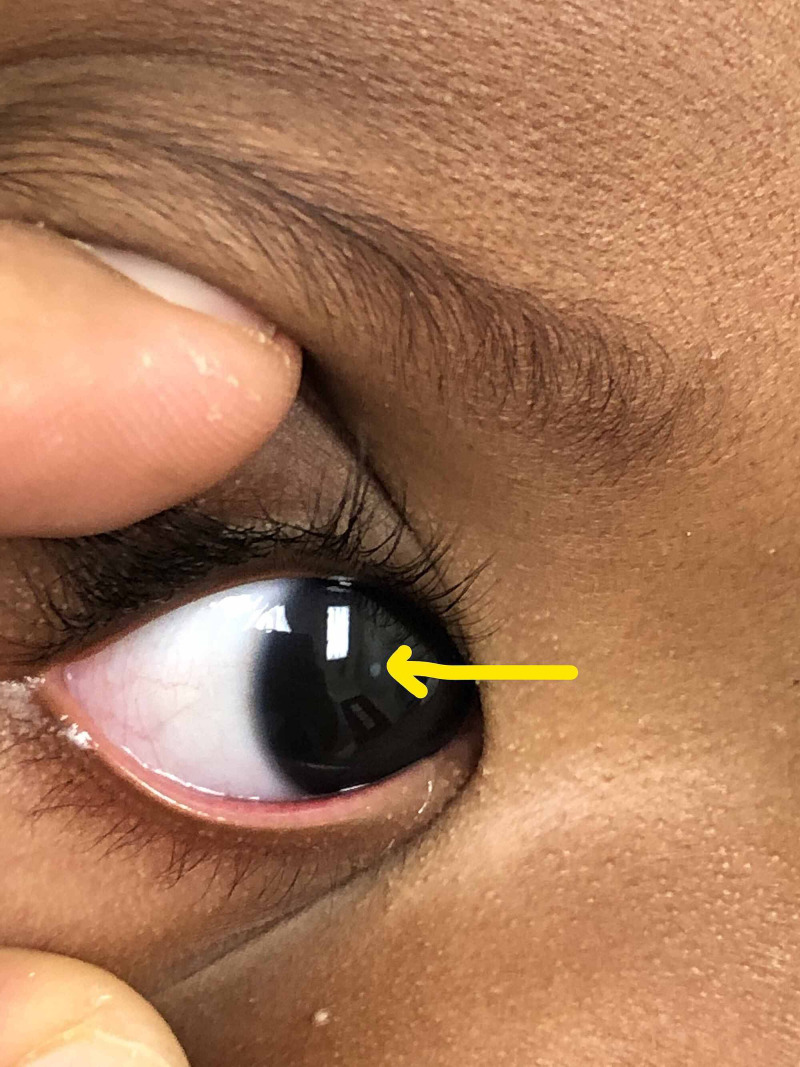
Why Leukocoria Happens
The normal red reflex results from light reflecting off the richly vascularized retina and choroid back through the pupil, and anything that opacifies or obstructs this pathway, whether a mass, a cataract, a retinal detachment, or a structural developmental anomaly, can replace that red glow with a white or yellow-white reflection instead.
Because so many structurally different conditions can each interrupt this same light pathway in their own way, the finding itself carries no specificity about cause, and the appearance of leukocoria on its own cannot distinguish a benign cataract from a malignant tumor without further evaluation.
This is precisely why leukocoria functions as a trigger for urgent, thorough evaluation rather than as a diagnosis that can be assessed casually at a routine visit.
Major Categories in the Differential
- Retinoblastoma, the most feared cause given its malignant potential and the urgency of prompt diagnosis and treatment
- Congenital cataract, discussed in its own dedicated article on this site, a common benign cause producing lens opacity that blocks the normal reflex
- Persistent fetal vasculature, discussed in its own dedicated article on this site, where remnant fetal vascular and fibrous tissue behind the lens produces a white reflex
- Coats disease, an exudative retinal vascular disorder that can produce a striking white or yellow subretinal exudative mass mimicking retinoblastoma closely
- Ocular toxocariasis, discussed in its own dedicated article on this site, an inflammatory granuloma from parasitic infection that is another classic retinoblastoma mimic
- Retinopathy of prematurity in its advanced stages, producing retinal detachment and fibrous tissue visible through the pupil in an appropriately premature infant
Fundus Explorer Pro
Photograph the retinal findings described here with the phone already in your pocket — 22 D optics and built-in illumination in one handheld unit.
From Choroida — the team behind this siteDistinguishing the Key Possibilities
Age at presentation, prematurity history, laterality, and family history each narrow the differential meaningfully: advanced retinopathy of prematurity requires a history of prematurity, Coats disease is classically unilateral in a young boy, and retinoblastoma has both sporadic and heritable forms with different associated risk patterns.
B-scan ultrasonography, discussed in its own dedicated article on this site, is a key early tool, since calcification, a common and relatively specific feature of retinoblastoma, is readily detected on ultrasound and helps distinguish it from most of its mimics.
MRI adds further information when ultrasound findings are ambiguous or when assessing for optic nerve or extraocular extension of a suspected tumor, information that is critical for staging and treatment planning if retinoblastoma is confirmed.
Why Urgency Matters
Any delay in correctly identifying retinoblastoma among the broader differential carries genuine risk, since this is a malignancy where prompt diagnosis and treatment materially affect both survival and the chance of preserving the eye and vision.
At the same time, a family understandably frightened by the possibility of cancer benefits from efficient, decisive evaluation that reaches a confident diagnosis, whether reassuring or serious, as quickly as possible rather than a prolonged, anxiety-inducing diagnostic process.
Referral to a center with pediatric ocular oncology expertise is appropriate whenever retinoblastoma cannot be confidently excluded on initial evaluation, given the specialized imaging and management this diagnosis, if confirmed, requires.
Practical Approach
A dilated fundus exam, ideally under sedation or anesthesia in a young child where cooperation is limited, remains the essential first step, often supplemented immediately by B-scan ultrasonography in the same setting.
Family history, systemic review for signs of prematurity-related or infectious causes, and careful attention to laterality and the specific character of the white reflex all contribute useful information that should be gathered systematically rather than skipped in the rush to obtain imaging.


Document what you see
Two smartphone imaging tools built for everyday clinic use — one for the slit lamp, one for the fundus.
From Choroida — the team behind this siteReferences
- Balmer A, Munier F. Differential diagnosis of leukocoria and strabismus, first presenting signs of retinoblastoma. Clinical Ophthalmology.
- American Academy of Ophthalmology. Basic and Clinical Science Course, Section 6: Pediatric Ophthalmology and Strabismus.
- Shields CL, Shields JA. Basic understanding of current classification and management of retinoblastoma. Current Opinion in Ophthalmology.
Test yourself
A few questions straight from this article.
-
Leukocoria, a white pupillary reflex in a child, is best understood as what?
Leukocoria is a sign, not a diagnosis, and its differential runs from benign cataract to life-threatening malignancy, so it triggers urgent systematic evaluation. -
The normal red reflex is produced by light reflecting off which structures?
Light reflects off the richly vascularised retina and choroid back through the pupil; anything opacifying that pathway replaces the red glow with a white reflection. -
Which cause of leukocoria is an exudative retinal vascular disorder, classically unilateral in a young boy?
Coats disease is an exudative retinal vascular disorder, classically unilateral in a young boy, producing a white or yellow subretinal exudative mass that closely mimics retinoblastoma. -
Which cause of leukocoria arises from remnant fetal vascular and fibrous tissue behind the lens?
In persistent fetal vasculature, remnant fetal vascular and fibrous tissue sitting behind the lens produces the white pupillary reflex. -
Which cause of leukocoria is an inflammatory granuloma resulting from parasitic infection?
Ocular toxocariasis produces an inflammatory granuloma from parasitic infection and is another classic retinoblastoma mimic. -
Which cause of leukocoria can only be invoked when there is a history of prematurity?
Advanced retinopathy of prematurity causes retinal detachment and fibrous tissue visible through the pupil, but requires an appropriately premature infant. -
Which B-scan ultrasound finding is common and relatively specific for retinoblastoma?
Calcification is a common and relatively specific feature of retinoblastoma, readily detected on ultrasound, which helps separate it from most of its mimics. -
What does MRI add beyond ultrasound in a child with suspected retinoblastoma?
MRI helps when ultrasound is ambiguous and assesses optic nerve or extraocular extension, information critical for staging and treatment planning. -
What is the essential first step in evaluating a young child with leukocoria?
A dilated fundus examination, ideally under sedation or anaesthesia where cooperation is limited, is the essential first step, often with B-scan ultrasound in the same setting. -
When should a child with leukocoria be referred for paediatric ocular oncology expertise?
Referral is appropriate whenever initial evaluation cannot confidently exclude retinoblastoma, given the specialised imaging and management it would require.

Writes and reviews the clinical articles on Educate. Every post is checked against peer-reviewed sources before it goes live.


