Juvenile idiopathic arthritis (JIA)-associated uveitis is a chronic, typically asymptomatic anterior uveitis that represents the single most common cause of uveitis in children.
Its silent nature is precisely what makes it dangerous: a substantial proportion of affected children have no eye symptoms at all until permanent structural damage has already occurred.
This is why screening protocols rather than symptom-triggered visits are the backbone of how this disease is actually caught.
Because the arthritis itself is usually diagnosed first, the ophthalmologist’s role here is fundamentally different from most other uveitis referrals: the child is examined not because of any eye complaint but purely because their rheumatologic diagnosis places them in a defined risk category for a disease they and their family may not even know to watch for.
Why It So Often Goes Unnoticed
Unlike most anterior uveitis in adults, JIA-associated uveitis is characteristically white and quiet.
The eye is not red, not painful, and the child has no complaints, even while active inflammation is present on slit-lamp exam.
This is the central clinical fact driving management of the disease: relying on the child or parent to report symptoms will miss most cases until they have already progressed, which is why regular, scheduled screening eye exams for at-risk children are considered standard of care rather than optional.
Risk Stratification
- Oligoarticular JIA (four or fewer joints involved) carries the highest uveitis risk, particularly in young girls with a positive antinuclear antibody (ANA)
- ANA-positive patients have a higher uveitis risk than ANA-negative patients across JIA subtypes
- Younger age at arthritis onset is associated with higher uveitis risk
- Systemic JIA and rheumatoid factor-positive polyarticular JIA carry comparatively lower uveitis risk
- Uveitis onset is usually within the first several years after arthritis diagnosis, though it can occasionally precede joint symptoms or develop after a longer interval
These risk factors are the basis for published screening interval guidelines (from bodies such as the American Academy of Pediatrics), which stratify how often a child should have a dilated slit-lamp exam based on JIA subtype, ANA status, and age at arthritis onset.
More frequent screening applies to higher-risk categories, tapering as time passes without uveitis developing or as the child ages out of the highest-risk window, though screening never stops entirely, since uveitis can still develop years after the initial arthritis diagnosis in some children.
All-fit Slit-Lamp Adapter
Record and share exactly what you see at the slit lamp. One adapter fits any slit lamp or surgical microscope — and any smartphone.
From Choroida — the team behind this siteExam Findings
- Anterior chamber cells and flare — the primary finding on which activity is graded, since the eye otherwise looks entirely normal externally
- Fine, non-granulomatous keratic precipitates in most cases
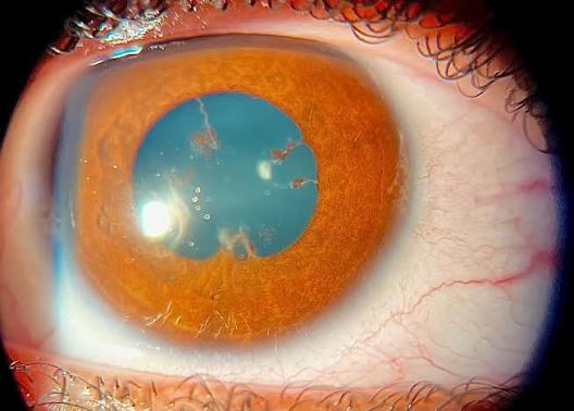
- Posterior synechiae, often the first sign a parent or clinician actually notices, since it can produce a visibly irregular pupil
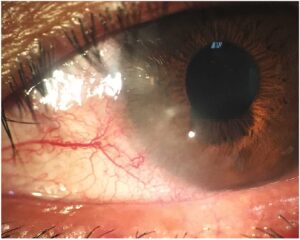
- Band keratopathy, from chronic calcium deposition in the setting of longstanding inflammation, discussed in more detail in this site’s dedicated article
- Cataract, frequently accelerated further by the corticosteroids commonly used to treat the underlying inflammation
- Glaucoma, from chronic inflammation itself or, again, from corticosteroid treatment — both mechanisms need to be considered when intraocular pressure is elevated in these patients
Complications From Delayed Diagnosis
Because the disease is silent, children are sometimes diagnosed only after band keratopathy, dense posterior synechiae, cataract, or glaucomatous damage has already developed — complications that are far harder to reverse than the underlying inflammation itself would have been if caught early.
This is the single strongest argument for adherence to structured screening protocols rather than waiting for a child to report visual symptoms, since by the time symptoms occur they often signal that significant, sometimes permanent, damage has already taken place.
Because visual outcomes correlate closely with how early treatment begins relative to the onset of inflammation, a missed or delayed screening visit is not simply a scheduling lapse — it represents a genuine, avoidable risk to the child’s long-term vision.
Management
Topical corticosteroids are first-line for controlling active inflammation, though chronic, frequent topical steroid use in a child carries its own substantial risk of cataract and glaucoma.
This is why steroid-sparing systemic therapy is introduced relatively early in children who need more than brief, low-dose topical treatment.
Methotrexate is typically the first steroid-sparing agent used, given its long track record of efficacy and the extensive pediatric rheumatology experience with its use in this population.
Biologic agents (TNF-alpha inhibitors such as adalimumab) have become increasingly important for children with disease that does not respond adequately to methotrexate alone, coordinated closely with pediatric rheumatology given the overlap between joint and eye disease management.
Regular monitoring for treatment-related complications (cataract, glaucoma) runs alongside monitoring for the underlying uveitis activity itself, since distinguishing disease-driven damage from treatment-related side effects is essential to adjusting the treatment plan appropriately.
Surgical management of cataract or band keratopathy, when needed, is generally deferred until inflammation is well controlled, because operating on an actively inflamed eye carries a substantially higher risk of poor outcomes and further inflammatory flare, so achieving a sustained quiet interval before surgery is typically a specific treatment goal in its own right.


Document what you see
Two smartphone imaging tools built for everyday clinic use — one for the slit lamp, one for the fundus.
From Choroida — the team behind this siteReferences
- American Academy of Pediatrics, Section on Rheumatology and Section on Ophthalmology. Guidelines for ophthalmologic examinations in children with juvenile rheumatoid arthritis. Pediatrics.
- Angeles-Han ST, Ringold S, Beukelman T, et al. American College of Rheumatology/Arthritis Foundation guideline for the screening, monitoring, and treatment of juvenile idiopathic arthritis-associated uveitis. Arthritis Care & Research.
- Heiligenhaus A, Michels H, Schumacher C, et al. Evidence-based, interdisciplinary guidelines for anti-inflammatory treatment of uveitis associated with juvenile idiopathic arthritis. Rheumatology International.
- American Academy of Ophthalmology. Basic and Clinical Science Course, Section 9: Intraocular Inflammation and Uveitis.
Test yourself
A few questions straight from this article.
-
Juvenile idiopathic arthritis-associated uveitis is best described as which pattern of intraocular inflammation?
It is a chronic anterior uveitis that is usually symptomless, and it is the single most common cause of uveitis in children. -
Which juvenile idiopathic arthritis subtype carries the highest risk of associated uveitis?
Oligoarticular disease, defined as four or fewer joints, carries the highest uveitis risk, particularly in young ANA-positive girls. Systemic and RF-positive polyarticular JIA carry comparatively lower risk. -
Which serologic result marks higher uveitis risk across juvenile idiopathic arthritis subtypes?
ANA-positive children have a higher uveitis risk than ANA-negative children, and ANA status is one of the variables used to set screening intervals. -
Why does juvenile idiopathic arthritis-associated uveitis usually escape detection without scheduled screening?
The eye is characteristically white and quiet with no complaints even while inflammation is active on slit lamp, so symptom-triggered visits miss most cases until damage has occurred. -
Which slit-lamp finding is the primary basis for grading activity in juvenile idiopathic arthritis-associated uveitis?
Since the eye looks entirely normal externally, anterior chamber cells and flare are the finding on which activity is actually graded. -
Keratic precipitates in juvenile idiopathic arthritis-associated uveitis are typically of which character?
Most cases show fine, non-granulomatous keratic precipitates rather than the granulomatous deposits seen in other uveitic entities. -
Which finding is often the first sign a parent or clinician actually notices in juvenile idiopathic arthritis-associated uveitis?
Because the eye is quiet, the visibly irregular pupil of posterior synechiae is frequently the first thing anyone notices. -
Which drug is typically the first steroid-sparing agent used in juvenile idiopathic arthritis-associated uveitis?
Methotrexate is usually first, given its long track record and the extensive paediatric rheumatology experience with it. TNF-alpha inhibitors such as adalimumab are added when methotrexate alone is inadequate. -
Why is steroid-sparing systemic therapy introduced relatively early in these children?
Topical corticosteroids are first-line for active inflammation, but sustained use in a child carries substantial cataract and glaucoma risk, so steroid-sparing therapy is started early. -
When is surgery for cataract or band keratopathy generally undertaken in these children?
Operating on an actively inflamed eye risks poor outcomes and further flare, so achieving a sustained quiet interval before surgery is itself a treatment goal.

Writes and reviews the clinical articles on Educate. Every post is checked against peer-reviewed sources before it goes live.