
Behçet uveitis is a relapsing, bilateral, nongranulomatous panuveitis with occlusive retinal vasculitis, and it is a leading cause of blindness among young men across the Mediterranean, Middle East, and Far East.
The eye may look white and comfortable between attacks, yet each relapse can add retinal ischemia and permanent damage.
Early systemic immunosuppression changes the outlook considerably.
Recognition depends on remembering the diagnosis in a young patient with recurrent uveitis and a history of oral ulcers.
What Is Behçet Disease?
Behçet disease is a systemic vasculitis affecting vessels of all sizes.
Its core features are recurrent oral aphthae, genital ulcers, skin lesions, and uveitis, and it may involve joints, vessels, and the central nervous system.
The International Study Group criteria (1990) require recurrent oral ulceration plus two of genital ulceration, eye lesions, skin lesions, or a positive pathergy test.
The newer International Criteria for Behçet Disease (2014) use a point system in which ocular lesions, oral aphthosis, and genital aphthosis each score two points, and a total of four or more supports the diagnosis.
The disease shows a strong association with HLA-B51, although HLA-B51 testing is not diagnostic on its own.
Ocular involvement is more frequent and more severe in young men.
Ocular Features of Behçet Uveitis
Anterior Segment
Anterior uveitis is often nongranulomatous and may include a hypopyon that forms without much redness, which is why the hypopyon may seem out of proportion to the symptoms.
The hypopyon shifts with head position and usually resolves quickly with treatment.
Posterior Segment
Posterior involvement determines the visual outcome.
Findings include:
- Vitritis, often marked during attacks
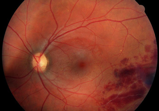
- Occlusive retinal vasculitis, mainly of veins, with sheathing and hemorrhages
- Retinal infiltrates that resolve rapidly in the acute phase
- Macular edema and ischemic maculopathy
- Optic disc hyperemia and later optic atrophy
- Retinal neovascularization and vitreous hemorrhage after ischemia
Fluorescein angiography shows diffuse capillary leakage with a fern-like pattern, disc leakage, and areas of nonperfusion.
Widefield angiography is useful because peripheral ischemia may be extensive.
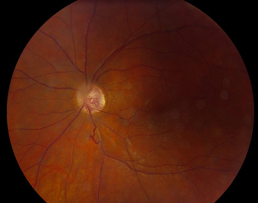
The course is episodic, and each attack may leave residual retinal thinning, vascular attenuation, and sclerosis.
Fundus Explorer Pro
Photograph the retinal findings described here with the phone already in your pocket — 22 D optics and built-in illumination in one handheld unit.
From Choroida — the team behind this siteDifferential Diagnosis
Other diseases that cause occlusive retinal vasculitis or recurrent panuveitis include:
- Tuberculosis (see ocular tuberculosis) and syphilis (see ocular syphilis)
- Sarcoidosis (see ocular sarcoidosis)
- Systemic lupus erythematosus and antiphospholipid syndrome
- Eales disease
- Multiple sclerosis
- HLA-B27-associated uveitis, which produces recurrent hypopyon
- Acute retinal necrosis and other viral retinitis
Behçet disease is a clinical diagnosis, and no single test confirms it.
Screening for tuberculosis and syphilis is justified before immunosuppression, particularly before starting a biologic agent.
Investigations
Fluorescein angiography is the key imaging test and should be repeated during follow-up, since retinal leakage may persist when the eye looks quiet.
Widefield angiography detects peripheral vascular leakage and nonperfusion that standard fields miss.
OCT identifies macular edema, epiretinal membrane, and outer retinal atrophy, and it tracks the response to treatment.
OCT angiography shows perifoveal capillary dropout in eyes with macular ischemia.
There are no specific laboratory tests, and a pathergy test is positive in only a proportion of patients, with variable frequency between regions.
The dermatologic and rheumatologic assessment supports the diagnosis and identifies extraocular disease that changes management.
Systemic Considerations
Vascular Behçet disease (venous thrombosis, arterial aneurysm) and neurologic involvement are the most serious extraocular manifestations.
Patients with retinal disease should have a low threshold for neurologic and vascular assessment, since ocular and systemic disease may coexist.
Male sex, young age at onset, and posterior segment disease predict a more severe course and justify early biologic therapy.
Women and older patients often have a milder course.
Pregnancy planning matters, because some immunosuppressive drugs are contraindicated, and the treatment plan should be reviewed before conception.
Treatment
Aims
The aims are to control attacks quickly, prevent relapse, and preserve the retina and macula.
Untreated posterior segment disease may cause blindness within a few years.
Corticosteroids
High-dose oral or intravenous corticosteroids control acute sight-threatening attacks.
Periocular or intravitreal steroids can help unilateral flares, and a dexamethasone implant may be considered for macular edema.
Steroid monotherapy is not enough for long-term control.
Conventional Immunosuppressants
The 2018 EULAR recommendations advise azathioprine together with systemic corticosteroids for Behçet patients with posterior segment involvement.
Cyclosporine A is an alternative, although it should be avoided when there is neurologic involvement.
Biologics
For severe disease, including retinal vasculitis or macular involvement, EULAR recommends infliximab or interferon alpha together with steroids.
Adalimumab is also used in practice.
Anti-TNF therapy can lower the frequency of attacks and improve visual outcome, but patients need screening for tuberculosis and hepatitis before starting.
Interferon alpha is effective, although side effects limit its use.
Local Complications
Panretinal photocoagulation is used for retinal neovascularization and severe ischemia.
Anti-VEGF injections may be used for macular edema or neovascularization in selected cases.
Vitrectomy is needed for vitreous hemorrhage that does not clear, tractional detachment, or cataract in the setting of quiescent disease.
Monitoring and Prognosis
Patients should be reviewed frequently in the first years, with fluorescein angiography and OCT used to detect silent activity.
Prognosis improved considerably with biologic therapy, although some eyes still develop optic atrophy, macular ischemia, and complicated cataract.
Systemic care is shared with rheumatology, and patients with neurologic or vascular symptoms need urgent assessment.


Document what you see
Two smartphone imaging tools built for everyday clinic use — one for the slit lamp, one for the fundus.
From Choroida — the team behind this siteReferences
- International Study Group for Behçet’s Disease. Criteria for diagnosis of Behçet’s disease. Lancet. 1990;335:1078-1080.
- International Team for the Revision of the International Criteria for Behçet’s Disease. The International Criteria for Behçet’s Disease (ICBD): a collaborative study of 27 countries on the sensitivity and specificity of the new criteria. J Eur Acad Dermatol Venereol. 2014;28:338-347.
- Hatemi G, Christensen R, Bang D, et al. 2018 update of the EULAR recommendations for the management of Behçet’s syndrome. Ann Rheum Dis. 2018;77:808-818.
- Tugal-Tutkun I, Onal S, Altan-Yaycioglu R, Huseyin Altunbas H, Urgancioglu M. Uveitis in Behçet disease: an analysis of 880 patients. Am J Ophthalmol. 2004;138:373-380.
- Yazici H, Seyahi E, Hatemi G, Yazici Y. Behçet syndrome: a contemporary view. Nat Rev Rheumatol. 2018;14:107-119.
Test yourself
A few questions straight from this article.
-
How is Behçet uveitis best characterised?
Behcet uveitis is a relapsing, bilateral, nongranulomatous panuveitis with occlusive retinal vasculitis, and a leading cause of blindness among young men in endemic regions. -
Under the 2014 International Criteria for Behçet Disease, what total score supports the diagnosis?
Ocular lesions, oral aphthosis and genital aphthosis each score two points, and a total of four or more supports the diagnosis. -
Which genetic marker shows a strong association with Behçet disease?
The association with HLA-B51 is strong, but the test is not diagnostic on its own, since Behcet disease remains a clinical diagnosis. -
What is characteristic of the hypopyon in Behçet anterior uveitis?
The hypopyon can appear out of proportion to the symptoms because it forms without much redness; it shifts with head position and usually clears quickly on treatment. -
Which fluorescein angiographic pattern is typical of Behçet uveitis?
Angiography shows diffuse capillary leakage in a fern-like pattern with disc leakage and areas of non-perfusion, and widefield imaging reveals how extensive peripheral ischaemia is. -
Why should fluorescein angiography be repeated during follow-up of Behçet uveitis?
The eye can look white and comfortable between attacks while leakage continues, so angiography and OCT are used to detect silent activity. -
Which features predict a more severe course of Behçet uveitis and justify early biologic therapy?
Male sex, young age at onset and posterior segment involvement all predict a worse course, while women and older patients often run a milder one. -
Which conventional immunosuppressant do the 2018 EULAR recommendations pair with steroids for posterior segment Behçet disease?
EULAR advises azathioprine with systemic corticosteroids for posterior segment involvement, and reserves infliximab or interferon alpha for severe retinal or macular disease. -
In which situation should cyclosporine A be avoided in Behçet disease?
Cyclosporine A is an alternative to azathioprine, but it should be avoided where there is neurologic involvement. -
Which other condition produces recurrent hypopyon uveitis and belongs in the differential with Behçet disease?
HLA-B27-associated uveitis also produces a recurrent hypopyon, which is why it sits in the differential alongside tuberculosis, syphilis, sarcoidosis and lupus.

Writes and reviews the clinical articles on Educate. Every post is checked against peer-reviewed sources before it goes live.


