Retinal neovascularization is the growth of abnormal new vessels from the retinal circulation onto the surface of the retina or the optic disc.
It is also the defining lesion of proliferative retinopathy.
It develops in response to ischemia, and it threatens vision through vitreous hemorrhage and traction.
Also, recognizing it early, and separating it from vessels that remain within the retina, drives the decision to treat.
What Is Retinal Neovascularization?
Areas of capillary nonperfusion release vascular endothelial growth factor and other angiogenic signals.
New vessels sprout from veins or capillaries, break through the internal limiting membrane, and grow along the posterior hyaloid face of the vitreous.
They have fenestrated, leaky walls and are accompanied by fibrous tissue, which contracts and pulls on the retina.
The vessels bleed easily, particularly when the vitreous moves, and the traction can cause retinal tears and detachment.
NVD and NVE
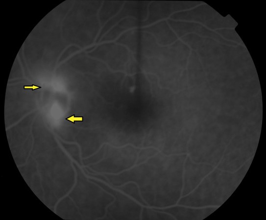
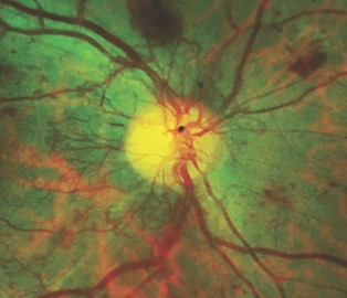
Doctors define neovascularization at the disc, or NVD, as new vessels on or within one disc diameter of the optic disc.
Neovascularization elsewhere, or NVE, lies farther out on the retina, usually along the vascular arcades at the border of nonperfused areas.
Also, NVD tends to carry a higher risk of vitreous hemorrhage than NVE of similar size, which is why the grading and the treatment thresholds differ.
Both forms can occur in the same eye, and new vessels can also form on the iris and in the angle (see rubeosis iridis).
Fundus Explorer Pro
Photograph the retinal findings described here with the phone already in your pocket — 22 D optics and built-in illumination in one handheld unit.
From Choroida — the team behind this siteCauses of Retinal Neovascularization
- Proliferative diabetic retinopathy: the commonest cause worldwide (see proliferative diabetic retinopathy)
- Retinal vein occlusion: especially ischemic central and branch occlusions
- Sickle cell retinopathy: sea fan neovascularization in the periphery (see sickle cell retinopathy)
- Retinopathy of prematurity
- Ocular ischemic syndrome and radiation retinopathy
- Inflammatory disease: Eales disease and some forms of retinal vasculitis (see Eales disease)
How to Recognize It
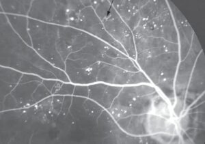
On examination, the vessels are fine, irregular, and arranged in a network or a frond.
They lie on the surface of the retina.
They cross over normal retinal vessels, which distinguishes them from intraretinal microvascular abnormalities (see intraretinal microvascular abnormalities).
Fluorescein angiography shows early, intense leakage that obscures the pattern in the late frames (see fluorescein angiography patterns).
In addition, OCT angiography shows flow above the internal limiting membrane, and widefield imaging reveals peripheral lesions that a standard view misses (see OCT angiography).
High-Risk Characteristics
The Diabetic Retinopathy Study defined features that predict severe visual loss without treatment.
- NVD of a quarter to a third of a disc area or more
- Any NVD with vitreous or preretinal hemorrhage
- NVE of at least half a disc area with vitreous or preretinal hemorrhage
Treat eyes with these features promptly, and in practice doctors also usually treat eyes with less severe neovascularization.
Treatment for Retinal Neovascularization
Panretinal photocoagulation has been the standard for decades.
It reduces the risk of severe visual loss by about half in high-risk eyes (see panretinal photocoagulation).
Also, intravitreal anti-VEGF drugs cause regression of neovascularization quickly, and trials have shown that ranibizumab was not inferior to laser for visual acuity at two years in proliferative diabetic retinopathy.
Anti-VEGF therapy requires repeated visits, because neovascularization returns when the injections stop.
Some patients drop out of follow-up (see anti-VEGF therapy).
Vitrectomy is also needed for nonclearing vitreous hemorrhage and for tractional detachment (see pars plana vitrectomy).
Treat the underlying disease as well, with systemic control of diabetes, blood pressure, and lipids, and treatment of any vein occlusion or inflammatory cause.
Follow-Up and Counselling
Review treated eyes at short intervals at first, to confirm regression and to look for recurrence.
Patients should also know that neovascularization can cause sudden floaters or loss of vision from bleeding, and that they should come in promptly.
Explain that treatment aims to protect the remaining vision and may not restore what has been lost.
Practical Examination Tips
Use a high-power lens at the slit lamp, such as a 78 or 90 diopter lens, and look for fine vessels at the edge of the disc and along the arcades.
Compare the vessels with a normal arteriole and venule to judge their caliber, and look for fibrous tissue that may also accompany them.
A widefield image in the clinic is useful, because clinicians can miss neovascularization elsewhere when they examine only the posterior pole.
Check the iris and angle without dilation in an eye with extensive neovascularization, because rubeosis may appear before the patient has symptoms.
Record the extent of neovascularization in disc areas.
The presence of fibrous tissue and hemorrhage, so that the response to treatment can be compared objectively at later visits.
For the latest studies, see the PubMed results for retinal neovascularization.


Document what you see
Two smartphone imaging tools built for everyday clinic use — one for the slit lamp, one for the fundus.
From Choroida — the team behind this siteReferences
- Diabetic Retinopathy Study Research Group. Photocoagulation treatment of proliferative diabetic retinopathy: clinical application of Diabetic Retinopathy Study (DRS) findings, DRS report number 8. Ophthalmology. 1981;88:583-600.
- Gross JG, Glassman AR, Jampol LM, et al. Panretinal photocoagulation vs intravitreous ranibizumab for proliferative diabetic retinopathy: a randomized clinical trial. JAMA. 2015;314:2137-2146.
- Ryan SJ, Schachat AP, Wilkinson CP, et al, eds. Retina. 6th ed. Elsevier; 2018.
- American Academy of Ophthalmology. Basic and Clinical Science Course, Section 12: Retina and Vitreous. AAO; current edition.

Writes and reviews the clinical articles on Educate. Every post is checked against peer-reviewed sources before it goes live.