
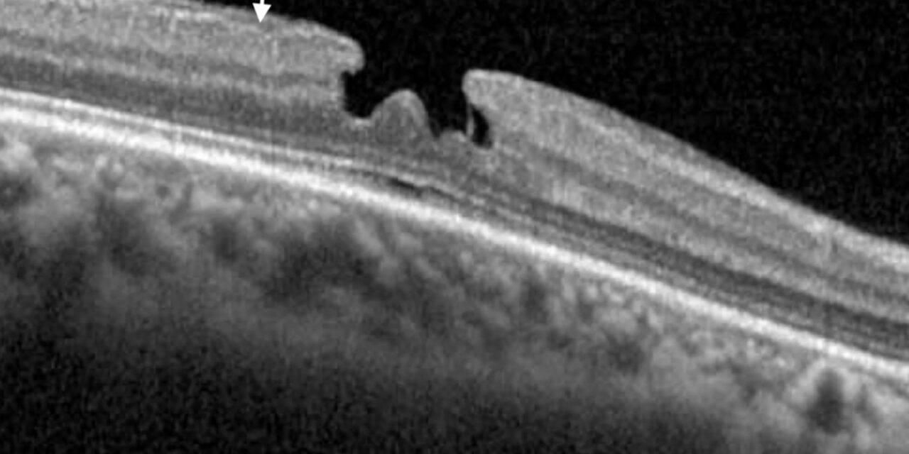
Lamellar versus full-thickness macular hole is a distinction that optical coherence tomography settled definitively, since these two entities can look deceptively similar on clinical examination alone but represent fundamentally different degrees of retinal tissue loss, with correspondingly different natural histories, surgical urgency, and expected outcomes.
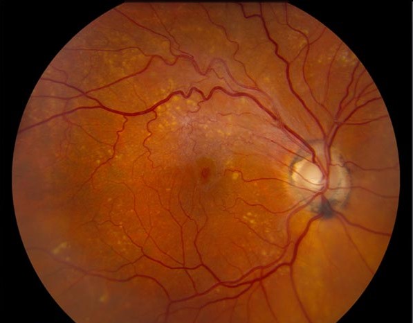
What Makes Them Fundamentally Different
A full-thickness macular hole involves a complete break spanning the entire thickness of the neurosensory retina at the fovea, from the internal limiting membrane through to the outer photoreceptor layer, typically resulting from anteroposterior and tangential vitreomacular traction pulling the foveal tissue apart.
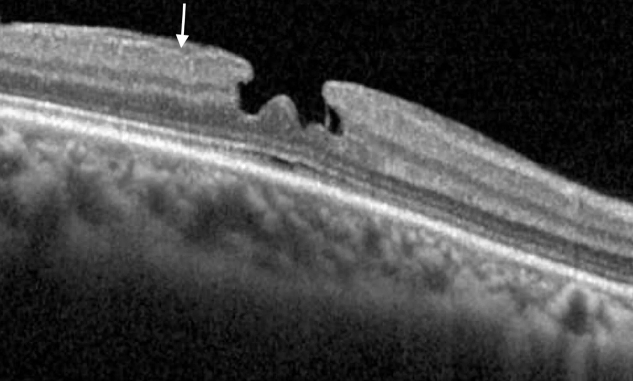
A lamellar macular hole, in contrast, involves only partial-thickness retinal tissue loss, typically affecting the inner retinal layers while the outer retinal layers, including the photoreceptor layer, remain intact, which is the central reason lamellar holes generally cause less severe visual impairment and behave so differently over time than their full-thickness counterparts.
Why OCT Is Essential for This Distinction
Before optical coherence tomography became routine, distinguishing these two entities reliably by clinical examination alone was genuinely difficult, and OCT’s ability to directly image the cross-sectional retinal architecture, clearly showing whether the outer retinal layers are intact or breached, is what made this an accurately and routinely distinguishable diagnosis in everyday practice.
Fundus Explorer Pro
Photograph the retinal findings described here with the phone already in your pocket — 22 D optics and built-in illumination in one handheld unit.
From Choroida — the team behind this siteFull-Thickness Macular Hole Features on OCT
- A complete, full-thickness retinal defect at the fovea, with no intact photoreceptor tissue bridging the base of the defect
- Often associated with a surrounding cuff of subretinal or intraretinal fluid and cystic changes in the adjacent retina
- Staged based on size and the presence or absence of a still-attached posterior hyaloid, which has direct implications for both natural history and surgical planning
Lamellar Macular Hole Features on OCT
- An irregular foveal contour with a partial-thickness defect, typically with an intact, continuous outer retinal layer at the base
- Often associated with epiretinal membrane, which can contribute a tangential traction component to the lamellar hole’s formation and progression
- Generally more stable over time than a full-thickness hole, with a lower likelihood of spontaneous progression to a full-thickness defect, though progression can occur in some cases
Macular Pseudohole: A Related but Distinct Entity
A macular pseudohole is yet another distinct entity, in which an epiretinal membrane with a central defect creates the clinical appearance of a hole without any actual retinal tissue loss at all, distinguished on OCT by a normal or even increased foveal retinal thickness, in contrast to the tissue thinning or loss characteristic of both lamellar and full-thickness holes.
Clinical and Visual Implications
- Full-thickness macular holes generally cause more significant central visual distortion and reduced visual acuity, given the complete disruption of foveal photoreceptor architecture
- Lamellar holes typically cause milder visual symptoms, sometimes primarily mild metamorphopsia or modest visual acuity reduction, consistent with the preserved outer retinal architecture
- Correctly distinguishing between the two guides realistic patient counseling about expected symptoms and the urgency, or lack thereof, of surgical intervention
Management
Full-Thickness Macular Hole
Surgical treatment, pars plana vitrectomy with internal limiting membrane peeling and gas tamponade, is the standard treatment for a full-thickness macular hole causing visual symptoms, generally recommended relatively promptly given evidence that earlier surgical intervention, before prolonged chronic hole duration, is associated with better anatomic and visual outcomes.
Lamellar Macular Hole
Management of lamellar holes is more variable and individualized, since many remain stable for years without significant progression or visual decline, supporting observation as a reasonable initial approach in many cases.
- Surgical intervention, generally vitrectomy with membrane peeling when a contributing epiretinal membrane is present, is considered for lamellar holes that are progressing, causing significant or worsening visual symptoms, or associated with substantial epiretinal membrane traction
- The decision to operate on a lamellar hole is generally less urgent and more individualized than for a full-thickness hole, reflecting the typically more indolent natural history of this condition
Prognosis
Full-thickness macular holes have a generally favorable surgical prognosis when treated with modern vitrectomy techniques, with high anatomic closure rates and meaningful visual improvement in most appropriately selected patients.
Lamellar macular holes have a good overall prognosis given their typically stable natural history, and even when surgery is eventually needed, outcomes are generally favorable, though the primary clinical value of correctly identifying a lamellar rather than full-thickness hole lies in avoiding unnecessary surgical urgency for a condition that, in many cases, simply does not require it.


Document what you see
Two smartphone imaging tools built for everyday clinic use — one for the slit lamp, one for the fundus.
From Choroida — the team behind this siteReferences
- Witkin AJ, Ko TH, Fujimoto JG, et al. Redefining lamellar holes and the vitreomacular interface: an ultrahigh-resolution optical coherence tomography study. Ophthalmology. 2006;113:388-397.
- Duker JS, Kaiser PK, Binder S, et al. The International Vitreomacular Traction Study Group classification of vitreomacular adhesion, traction, and macular hole. Ophthalmology. 2013;120:2611-2619.
- Govetto A, Dacquay Y, Farajzadeh M, et al. Lamellar macular hole: two distinct clinical entities? Am J Ophthalmol. 2016;164:99-109.
- Kelly NE, Wendel RT. Vitreous surgery for idiopathic macular holes. Results of a pilot study. Arch Ophthalmol. 1991;109:654-659.
