
This guide is now part of our complete overview: Eales Disease: Diagnosis, Differential and Treatment.
Eales disease is an idiopathic peripheral retinal vasculitis, typically affecting young, otherwise healthy adult men, characterized by peripheral retinal vascular inflammation and occlusion that predisposes to recurrent vitreous hemorrhage.
It remains a diagnosis of exclusion, made only after a careful search for the many recognized causes of retinal vasculitis and peripheral vascular occlusion has failed to identify a specific underlying etiology, despite decades of study into a possible infectious or immunologic trigger.
Because it is defined partly by what it is not, a systematic and thorough exclusion of mimicking conditions is inseparable from actually making the diagnosis correctly.
The disease shows a notably higher reported incidence in certain regions, particularly parts of South Asia, than elsewhere, a geographic pattern that has fueled much of the long-standing interest in an infectious or environmental trigger.
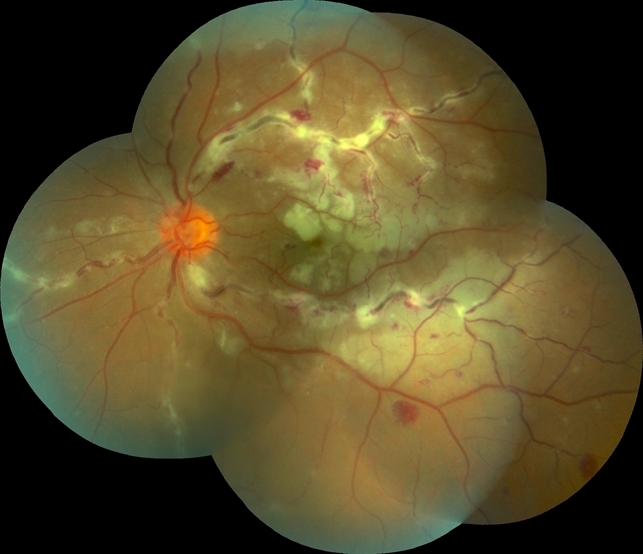
Clinical Course
The disease classically progresses through overlapping stages: an initial phase of peripheral retinal vasculitis, most often periphlebitis, followed by peripheral vascular non-perfusion, and eventually neovascularization arising from the areas of ischemia.
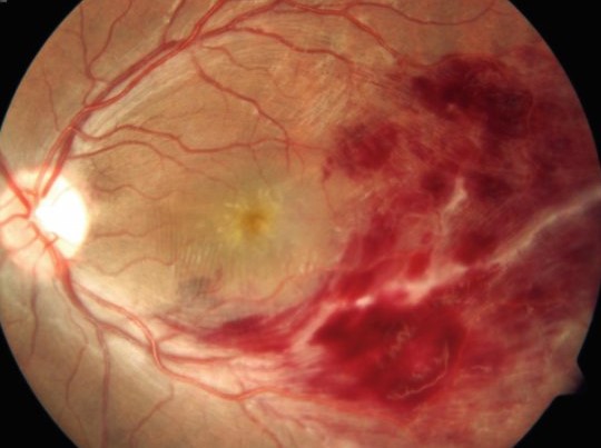
Once neovascular vessels form, they are fragile and prone to bleeding into the vitreous, which is what produces the recurrent vitreous hemorrhage that is often the presenting complaint bringing a young, previously asymptomatic patient to medical attention.
The disease is frequently bilateral, though the two eyes can be affected asymmetrically and at different points in their individual disease course, meaning one eye may be actively hemorrhaging while the other shows only early, asymptomatic peripheral vasculitis.
Clinical Presentation
- Sudden painless vision loss or floaters from vitreous hemorrhage, often the presenting complaint in a young, healthy adult male with no prior ocular history
- Peripheral retinal vascular sheathing and inflammation on exam of the fellow or less severely affected eye, when the view allows adequate visualization
- Peripheral neovascularization, best appreciated on fluorescein angiography, arising at the border of perfused and non-perfused peripheral retina
- A demographic pattern strongly skewed toward young adult men, typically in their twenties or thirties, a distinctive epidemiologic clue that helps distinguish Eales disease from many of its mimics
Fundus Explorer Pro
Photograph the retinal findings described here with the phone already in your pocket — 22 D optics and built-in illumination in one handheld unit.
From Choroida — the team behind this siteDifferential Diagnosis
Diabetic retinopathy can produce a broadly similar picture of peripheral non-perfusion and neovascularization, but typically occurs in an older patient with an established diabetes history and a different, more central pattern of vascular change.
Retinal vein occlusion, sickle cell retinopathy, and sarcoidosis-associated retinal vasculitis, discussed in relation to ocular sarcoidosis in its own dedicated article on this site, all need to be actively excluded given their overlapping potential to produce peripheral vasculitis and non-perfusion.
Infectious causes, particularly tuberculosis, have long been proposed as a possible trigger for Eales disease specifically, and a history and workup for latent or active tuberculosis exposure is a standard part of the evaluation in endemic regions, even though the exact causal relationship remains unproven.
Diagnostic Evaluation
Fluorescein angiography is essential for mapping the extent of peripheral non-perfusion and identifying areas of active or subclinical neovascularization, guiding both diagnosis and subsequent laser treatment planning.
A thorough systemic workup, including screening for diabetes, sickle cell disease, sarcoidosis, tuberculosis, and other systemic vasculitides, is necessary to support the diagnosis of exclusion, since Eales disease cannot be confidently diagnosed until these other causes have been reasonably ruled out.
B-scan ultrasonography, discussed in its own dedicated article on this site, is useful when vitreous hemorrhage precludes an adequate direct view of the retina, helping assess for an underlying retinal detachment that would change the urgency and approach to treatment.
Management
Peripheral scatter laser photocoagulation is applied to areas of retinal non-perfusion and neovascularization, following the same general treatment logic used for other ischemic retinal diseases, aiming to reduce the angiogenic drive behind ongoing neovascularization and hemorrhage risk.
Corticosteroids or other immunosuppressive therapy may be used during the active vasculitic phase to control inflammation, particularly when active periphlebitis is present alongside the ischemic and neovascular findings.
Vitrectomy is reserved for eyes with non-clearing vitreous hemorrhage, tractional retinal detachment, or other complications that do not resolve with laser and medical management alone, following similar surgical principles to other proliferative retinal vascular diseases.
Because both eyes are frequently involved even when only one is currently symptomatic, careful examination and monitoring of the fellow eye is a standard, essential part of managing every confirmed case.


Document what you see
Two smartphone imaging tools built for everyday clinic use — one for the slit lamp, one for the fundus.
From Choroida — the team behind this siteReferences
- Biswas J, Sharma T, Gopal L, et al. Eales disease: an update. Survey of Ophthalmology.
- American Academy of Ophthalmology. Basic and Clinical Science Course, Section 12: Retina and Vitreous.
- Saxena S, Kumar D. Eales disease: an update. Journal of Postgraduate Medicine.
Test yourself
A few questions straight from this article.
-
What must happen before Eales disease can be diagnosed?
Eales disease is idiopathic and remains a diagnosis of exclusion, reached only after a systematic search for recognised causes of retinal vasculitis and vascular occlusion comes up empty. -
Eales disease characteristically affects which group of patients?
The demographic skew toward otherwise healthy young adult men in their twenties or thirties is a distinctive epidemiologic clue separating it from many mimics. -
Which sequence describes the classic progression of Eales disease?
The disease moves through overlapping stages: peripheral vasculitis, usually periphlebitis, then peripheral vascular non-perfusion, then neovascularisation from the ischaemic retina. -
Why is examination and monitoring of the fellow eye essential in every confirmed Eales case?
Involvement is frequently bilateral but asymmetric, so one eye may be haemorrhaging while the other shows only early, asymptomatic peripheral vasculitis. -
Which feature distinguishes diabetic retinopathy from Eales disease?
Diabetic retinopathy can look broadly similar but occurs in an older patient with a known diabetes history and a different, more central pattern of vascular change. -
Which infectious agent has long been proposed as a possible trigger for Eales disease?
Tuberculosis has long been proposed as a trigger, and workup for latent or active exposure is standard in endemic regions, although the causal relationship remains unproven. -
What is fluorescein angiography essential for in Eales disease?
Angiography defines the extent of peripheral non-perfusion and finds active or subclinical new vessels, guiding both the diagnosis and the laser treatment plan. -
When is B-scan ultrasonography particularly useful in Eales disease?
With the fundus obscured by haemorrhage, ultrasound assesses for an underlying retinal detachment, which changes both the urgency and the approach to treatment. -
Vitrectomy in Eales disease is reserved for which situation?
Surgery is held back for eyes with non-clearing vitreous haemorrhage, tractional retinal detachment or other complications that laser and medical treatment do not resolve. -
Peripheral scatter laser photocoagulation in Eales disease is applied to what?
Laser is directed at non-perfused retina and neovascularisation to reduce the angiogenic drive behind continuing new vessel growth and bleeding.

Writes and reviews the clinical articles on Educate. Every post is checked against peer-reviewed sources before it goes live.


