
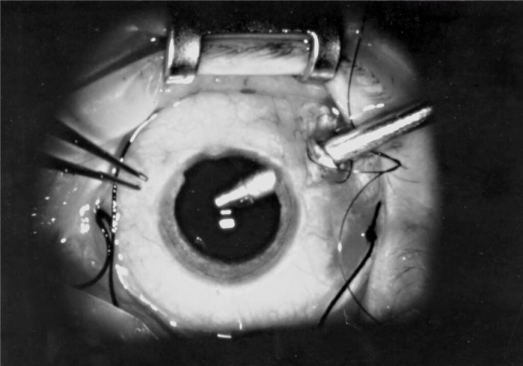
Vitreoretinal surgery began in 1970 with an instrument the size of a pen that could cut and aspirate vitreous through a single small opening in the eye wall.
Everything about modern retinal surgery – macular hole repair, retinal detachment reattachment, diabetic vitreous haemorrhage clearance – descends from that one idea.
Pars plana vitrectomy (PPV) is the platform procedure of retinal surgery, and the story of its evolution is largely a story of instruments becoming smaller.
What started as a 20-gauge procedure requiring sutured wounds is now routinely performed through 25 or even 27-gauge ports that self-seal.
Understanding what the surgery actually does – and where it stops being enough on its own – is essential to understanding almost every other retinal treatment covered on this site.
What Is Pars Plana Vitrectomy?
Pars plana vitrectomy is removal of the vitreous gel through small incisions in the pars plana, a region of the eye wall that can be entered safely without damaging the retina or lens.
It serves two distinct surgical purposes:
- Removing vitreous that is itself the problem – haemorrhage, opacity, infection, or traction pulling on the retina
- Creating surgical access to the retina and macula for procedures that could not otherwise be performed – membrane peeling, retinal reattachment, or delivery of subretinal therapy
In practice, most vitrectomies do both at once: the vitreous is cleared largely to allow safe, controlled work on the retina behind it.
Indications
Vitreous-Primary Disease
- Non-clearing vitreous haemorrhage, commonly from proliferative diabetic retinopathy
- Endophthalmitis, where vitrectomy both reduces infective and inflammatory load and allows targeted intravitreal antibiotic delivery
- Vitreous opacities significantly affecting vision
Retinal Disease Requiring Surgical Access
- Rhegmatogenous retinal detachment, particularly complex or posterior detachments unsuited to scleral buckling alone
- Macular hole, requiring internal limiting membrane peeling
- Epiretinal membrane causing visually significant distortion
- Proliferative diabetic retinopathy with tractional retinal detachment
- Dislocated intraocular lens or retained lens fragments after complicated cataract surgery
- Subretinal gene therapy delivery, which requires vitrectomy as the access route to the subretinal space
That last indication is a genuinely new category – vitrectomy has become the delivery platform for a whole class of therapies that did not exist a decade ago.
Fundus Explorer Pro
Photograph the retinal findings described here with the phone already in your pocket — 22 D optics and built-in illumination in one handheld unit.
From Choroida — the team behind this siteSurgical Technique
Access
- Three or more small sclerotomies are made through the pars plana, positioned to avoid the lens anteriorly and the retina posteriorly
- Modern small-gauge systems (23, 25 or 27-gauge) use self-sealing, sutureless incisions in most cases, reducing surgical trauma and speeding recovery compared with older 20-gauge sutured surgery
- Ports are used for the infusion line, the light source, and the working instrument
Core Steps
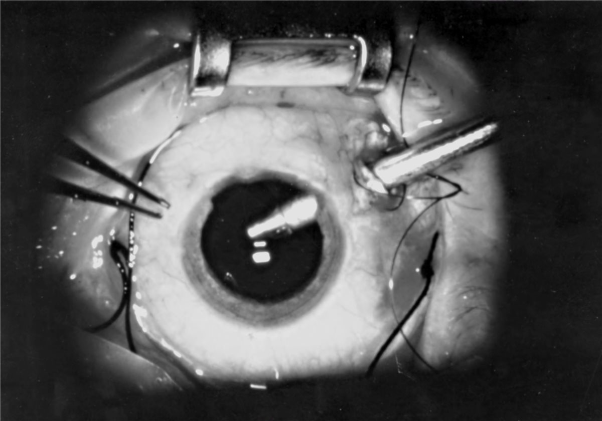
- The vitreous cutter removes the vitreous gel while the infusion line maintains intraocular pressure and volume
- Posterior vitreous detachment is induced if not already present, since residual vitreous cortex is a major driver of postoperative traction and re-detachment
- Additional manoeuvres are performed depending on the indication – membrane peeling with intraocular forceps, laser retinopexy around retinal breaks, or drainage of subretinal fluid
- An endotamponade agent – gas or, less commonly, silicone oil – may be placed at the end of surgery to support the retina while it heals
Closure
- Sclerotomies are checked for leakage and sutured only if they fail to self-seal
- Postoperative positioning instructions are given if a gas or oil tamponade has been used, since gravity determines where the bubble supports the retina
The move to sutureless small-gauge surgery is arguably the single biggest advance in this field over the last two decades – it converted a procedure with days of discomfort into one with next-day recovery for many patients.
Endotamponade Agents
- Air – short-acting, used for straightforward cases needing only brief support
- Gas – sulfur hexafluoride or perfluoropropane, providing intermediate to long-acting tamponade that resorbs over weeks; requires strict avoidance of air travel and nitrous oxide anaesthesia until fully absorbed
- Silicone oil – long-term tamponade for complex or recurrent detachments, requiring a planned second procedure for removal and carrying its own risk of emulsification and secondary glaucoma
The choice of tamponade is a judgment about how long mechanical support is needed to let the retina reattach and heal – it is a means to an end, not the definitive treatment itself.
Complications
- Cataract progression – the single most common long-term consequence of vitrectomy in phakic eyes, occurring in the great majority of patients within a few years
- Retinal detachment or new retinal breaks, from traction created during vitreous removal or membrane peeling
- Elevated intraocular pressure, particularly with gas or silicone oil tamponade
- Endophthalmitis – rare but sight-threatening, as with any intraocular surgery
- Silicone oil emulsification, migration into the anterior chamber, and oil-related glaucoma with prolonged tamponade
- Recurrent vitreous haemorrhage, particularly in diabetic eyes with residual ischaemic drive
Cataract is worth flagging specifically to patients before surgery, since it is near-universal in phakic eyes and often the first thing patients notice once their primary retinal problem has resolved.
Prognosis
Outcomes depend heavily on the underlying indication rather than on the vitrectomy technique itself.
- Macular hole surgery achieves anatomical closure in the large majority of appropriately selected cases, with good visual recovery when performed before chronic photoreceptor loss
- Retinal detachment repair achieves single-surgery anatomical success in most primary cases, though final visual outcome depends heavily on whether the macula was detached preoperatively and for how long
- Vitreous haemorrhage clearance in diabetic eyes restores vision effectively, but long-term outcome still depends on control of the underlying retinopathy
- Complex tractional and combined detachments carry a lower single-surgery success rate and a higher rate of further intervention
Because vitrectomy is a platform rather than a single disease treatment, its “prognosis” is really the prognosis of whatever it was performed to fix – the surgery succeeds when it enables the right definitive step, not in isolation.


Document what you see
Two smartphone imaging tools built for everyday clinic use — one for the slit lamp, one for the fundus.
From Choroida — the team behind this siteReferences
- Machemer R, Buettner H, Norton EW, Parel JM. Vitrectomy: a pars plana approach. Transactions of the American Academy of Ophthalmology and Otolaryngology. 1971.
- Fujii GY, De Juan E, Humayun MS, et al. A new 25-gauge instrument system for transconjunctival sutureless vitrectomy surgery. Ophthalmology. 2002.
- American Academy of Ophthalmology. Vitreoretinal Surgery Preferred Practice Pattern.
- Pars Plana Vitrectomy. EyeWiki, American Academy of Ophthalmology.
- Vitrectomy. StatPearls, NCBI Bookshelf.
Test yourself
A few questions straight from this article.
-
Why are the incisions for vitrectomy placed at the pars plana?
The pars plana is a region of the eye wall that can be entered safely, allowing instruments in without damaging the retina posteriorly or the lens anteriorly. -
Which additional manoeuvre does macular hole repair require during pars plana vitrectomy?
Macular hole is listed among the retinal conditions requiring surgical access, with internal limiting membrane peeling as the manoeuvre that makes closure possible. -
What is the main advantage of modern 23, 25 and 27-gauge vitrectomy over older 20-gauge surgery?
Small-gauge systems use self-sealing, sutureless incisions in most cases, reducing surgical trauma and speeding recovery compared with older sutured 20-gauge surgery. -
Why is a posterior vitreous detachment induced during vitrectomy when one is not already present?
Residual vitreous cortex left on the retina is a major driver of postoperative traction and re-detachment, so a posterior vitreous detachment is induced if absent. -
What are the two benefits of vitrectomy in endophthalmitis?
In endophthalmitis, vitrectomy both reduces the infective and inflammatory load and provides a route for targeted intravitreal antibiotic delivery. -
A patient with intraocular gas tamponade after vitrectomy must avoid which two things until it absorbs?
Sulfur hexafluoride and perfluoropropane tamponade require strict avoidance of air travel and nitrous oxide anaesthesia until the bubble is fully absorbed. -
Which burden distinguishes silicone oil from gas as an endotamponade after vitrectomy?
Silicone oil gives long-term tamponade for complex or recurrent detachments but requires a planned second operation to remove it, and risks emulsification and secondary glaucoma. -
What is the most common long-term consequence of pars plana vitrectomy in a phakic eye?
Cataract progression is the single most common long-term consequence of vitrectomy in phakic eyes, occurring in the great majority of patients within a few years. -
After vitrectomy for rhegmatogenous retinal detachment, what most determines the final visual outcome?
Single-surgery anatomical success is achieved in most primary cases, but final vision depends heavily on whether the macula was detached before surgery and for how long. -
Why is pars plana vitrectomy needed before subretinal gene therapy can be delivered?
Subretinal gene therapy delivery requires vitrectomy as the access route to the subretinal space, making the surgery a platform for treatments that did not exist a decade ago.

Writes and reviews the clinical articles on Educate. Every post is checked against peer-reviewed sources before it goes live.


