
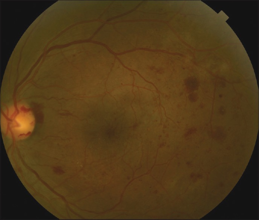
Vitreous hemorrhage in diabetes is most often the first visible sign that proliferative retinopathy has crossed from a silent process into one that threatens vision suddenly, and the workup has to answer two questions at once: how bad is the bleeding source, and is the retina underneath still attached.
Both questions matter for deciding whether to wait or operate.
Why Diabetic Vitreous Hemorrhage Happens
Retinal ischemia in diabetic retinopathy drives new vessel growth along the posterior hyaloid, and these fragile new vessels lack the structural integrity of normal retinal vessels.
Vitreous traction on the posterior hyaloid, particularly during a posterior vitreous detachment, tears these vessels and causes bleeding into the vitreous cavity, sometimes precipitated by a Valsalva-type event such as coughing or straining, though often occurring without any identifiable trigger.
Clinical Presentation
- Sudden onset of floaters, ranging from a few new spots to a dense shower
- Blurred or significantly reduced vision, depending on the amount of blood
- A red or absent fundus reflex on examination when hemorrhage is dense
- Symptoms are sometimes preceded by a period of flashing lights if an acute posterior vitreous detachment is the trigger
Fundus Explorer Pro
Photograph the retinal findings described here with the phone already in your pocket — 22 D optics and built-in illumination in one handheld unit.
From Choroida — the team behind this siteEvaluation
History and Examination
A history of diabetic retinopathy severity, prior laser treatment, and glycemic control helps frame the likely extent of underlying disease.
Examination assesses what can be seen of the fundus, however limited, along with the fellow eye, which often shows the stage of retinopathy more clearly when the affected eye’s view is obscured.
B-Scan Ultrasonography
When the fundus view is significantly obscured, B-scan ultrasonography is the key test, assessing for retinal detachment, the extent of vitreous hemorrhage, and any posterior vitreous detachment.
Specific attention is paid to whether the retina appears attached, since a tractional or combined tractional-rhegmatogenous detachment underlying the hemorrhage changes the urgency and type of surgery needed (see combined rhegmatogenous-tractional retinal detachment).
Fellow Eye Assessment
The fellow eye is examined in detail, since its retinopathy severity, presence of proliferative disease, and prior treatment provide important context and sometimes prompt treatment of that eye even while the affected eye is obscured.
Management
Observation
Many episodes of vitreous hemorrhage are managed initially with observation, allowing time for spontaneous clearing, particularly when B-scan confirms an attached retina and there is no urgent indication for surgery.
Patients are advised to keep the head elevated, which can help blood settle inferiorly, and to avoid anticoagulant use where medically appropriate and avoid strenuous activity that might provoke rebleeding.
Panretinal Photocoagulation
When some view is available, or once hemorrhage clears enough, prompt panretinal photocoagulation to the treatable retina reduces the drive for further neovascularization and lowers the risk of recurrent bleeding (see panretinal photocoagulation).
Anti-VEGF Therapy
Anti-VEGF injection can be used to reduce neovascular activity and has been used to help clear vitreous hemorrhage or to reduce bleeding risk before planned vitrectomy, though it is not a substitute for definitive treatment of the underlying ischemic retina.
Pars Plana Vitrectomy
Surgery is indicated for nonclearing vitreous hemorrhage after an appropriate observation period, for hemorrhage obscuring the view in an eye with suspected tractional retinal detachment threatening the macula, for dense premacular hemorrhage, or for bilateral significant hemorrhage where visual function is severely limited (see pars plana vitrectomy and proliferative diabetic retinopathy).
Timing is individualized: a young patient with type 1 diabetes and dense hemorrhage, in whom spontaneous clearing is less likely and the risk of progression to tractional detachment is higher, is generally offered earlier surgery than an older patient with a first, milder episode.
Prognosis
Many episodes clear with observation and appropriate laser treatment once the view improves, particularly in eyes without significant traction.
The presence of tractional retinal detachment at presentation, poor glycemic and blood pressure control, and delayed treatment of the underlying proliferative disease are associated with worse visual outcomes.


Document what you see
Two smartphone imaging tools built for everyday clinic use — one for the slit lamp, one for the fundus.
From Choroida — the team behind this siteReferences
- Diabetic Retinopathy Clinical Research Network. Early vitrectomy for vitreous hemorrhage from proliferative diabetic retinopathy: two-year results of a randomized trial. Ophthalmology. 2010;117:1087-1093.
- The Diabetic Retinopathy Vitrectomy Study Research Group. Early vitrectomy for severe vitreous hemorrhage in diabetic retinopathy. Two-year results of a randomized trial. Arch Ophthalmol. 1985;103:1644-1652.
- Faghihi H, Taheri A, Farahvash MS, Esfahani MR, Rajabi MT. Fundus microperimetry in eyes with vitreous hemorrhage. J Ophthalmic Vis Res. 2009;4:75-79.
- Sarrafizadeh R, Hassan TS, Ruby AJ, et al. Incidence of retinal detachment and visual outcome in eyes presenting with spontaneous vitreous hemorrhage. Ophthalmology. 2001;108:2273-2278.
Test yourself
A few questions straight from this article.
-
What mechanism causes vitreous hemorrhage in proliferative diabetic retinopathy?
Ischaemia drives fragile new vessels along the posterior hyaloid, and traction on that hyaloid, especially during posterior vitreous detachment, tears them. -
Which examination finding suggests dense vitreous hemorrhage in a diabetic eye?
When the haemorrhage is dense the fundus reflex becomes red or disappears altogether, limiting any view of the retina behind it. -
When the fundus view is significantly obscured by diabetic vitreous hemorrhage, which test is key?
B-scan assesses the extent of haemorrhage, the presence of posterior vitreous detachment, and above all whether the retina is detached. -
What must B-scan ultrasonography establish in a diabetic eye with vitreous hemorrhage?
A tractional or combined tractional-rhegmatogenous detachment under the blood changes both the urgency and the type of surgery required. -
Why is the fellow eye examined in detail in diabetic vitreous hemorrhage?
The fellow eye often shows the stage of retinopathy more clearly, and its own proliferative disease may warrant treatment while the affected eye stays obscured. -
Which positional advice is given during observation of vitreous hemorrhage?
An elevated head position helps blood settle inferiorly, improving the view while spontaneous clearing is awaited. -
What does panretinal photocoagulation achieve in diabetic vitreous hemorrhage?
Treating the accessible retina reduces the stimulus for further neovascularisation and lowers the risk of recurrent bleeding. -
What is the role of anti-VEGF therapy in diabetic vitreous hemorrhage?
Anti-VEGF can reduce neovascular activity and bleeding risk before vitrectomy, but it does not substitute for definitive treatment of the ischaemic retina. -
Which patient with diabetic vitreous hemorrhage is generally offered earlier vitrectomy?
Spontaneous clearing is less likely and progression to tractional detachment more likely in such eyes, so surgery is offered sooner. -
Which factors are associated with worse visual outcomes after diabetic vitreous hemorrhage?
Traction at presentation, poor glycaemic and blood pressure control, and delayed treatment of the proliferative disease all predict worse vision.

Writes and reviews the clinical articles on Educate. Every post is checked against peer-reviewed sources before it goes live.


