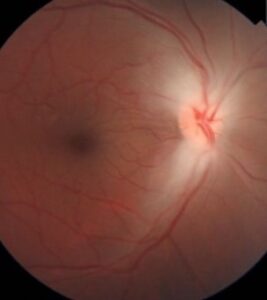
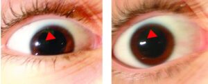
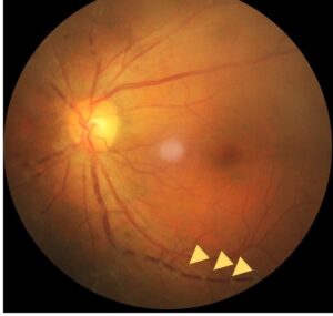
Horner syndrome is the classic triad of ptosis, miosis, and anhidrosis resulting from interruption anywhere along the three-neuron oculosympathetic pathway.
Its diagnostic value lies less in the triad itself — which is often subtle and easy to miss — than in the localizing workup that follows once it is recognized.
This is because the pathway’s long, three-neuron course means the underlying cause can be located anywhere from the hypothalamus down to the neck, with different implications depending on where along that path the interruption sits.
The Three-Neuron Pathway
- First-order (central) neuron — runs from the hypothalamus through the brainstem to the ciliospinal center of Budge in the spinal cord (roughly C8–T2)
- Second-order (preganglionic) neuron — exits the spinal cord, passes near the apex of the lung, and ascends through the neck to the superior cervical ganglion
- Third-order (postganglionic) neuron — travels from the superior cervical ganglion along the internal carotid artery, through the cavernous sinus, and finally reaches the eye
A lesion anywhere along this entire pathway produces the same basic clinical triad, which is precisely why localization matters so much.
First-order lesions raise concern for brainstem or spinal cord pathology, second-order lesions raise concern for apical lung tumor (Pancoast tumor) or other neck/chest pathology, and third-order lesions raise concern for carotid artery dissection, among other causes specific to that segment.
Clinical Findings
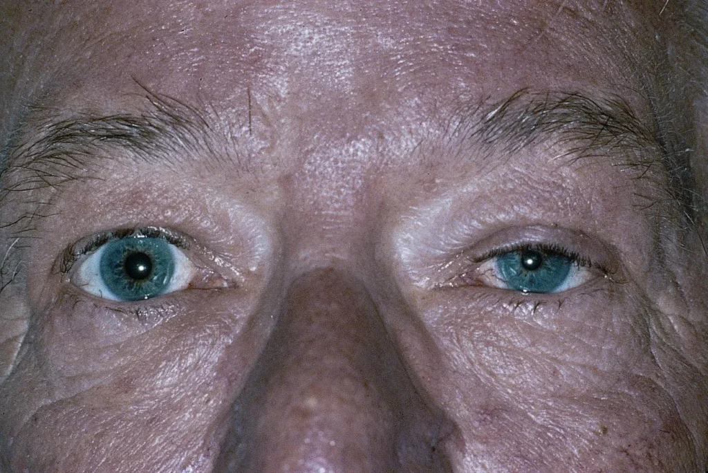
- Mild ptosis (typically 1–2 mm, from loss of Müller’s muscle function, distinctly less than the ptosis of a third nerve palsy) and, sometimes, a subtle “upside-down ptosis” — slight elevation of the lower lid from loss of the analogous smooth muscle there
- Miosis — the affected pupil is smaller than the normal side, and the anisocoria is characteristically greater in dim light (when the normal pupil dilates well and the affected one cannot)
- Dilation lag — the affected pupil dilates more slowly than the normal pupil when lights are dimmed, a subtle but useful sign distinct from simply comparing final pupil sizes
- Anhidrosis — reduced sweating, present only with lesions proximal to where the sudomotor fibers diverge from the pathway (roughly at the level of the carotid bifurcation), which is why anhidrosis is a feature of first- and second-order lesions but often absent in third-order (postganglionic) lesions
- Iris heterochromia — the affected iris may be lighter in congenital or longstanding Horner syndrome, from a lifelong lack of sympathetic trophic input to melanocyte development, and is a specific clue toward a congenital rather than acute acquired cause
All-fit Slit-Lamp Adapter
Record and share exactly what you see at the slit lamp. One adapter fits any slit lamp or surgical microscope — and any smartphone.
From Choroida — the team behind this siteCauses by Level
- First-order — brainstem stroke (lateral medullary/Wallenberg syndrome being a classic cause), demyelinating disease, spinal cord tumor or syrinx
- Second-order — apical lung tumor (Pancoast tumor), a cause specifically worth remembering and actively excluding; thyroid malignancy; trauma or surgery in the neck or chest; central line placement or chest tube complications
- Third-order — internal carotid artery dissection, a genuine emergency given the associated stroke risk, classically presenting with acute, painful Horner syndrome, sometimes with ipsilateral neck or face pain; cavernous sinus pathology; cluster headache, during which a transient, painful Horner syndrome can occur
- Congenital — often related to birth trauma (particularly with brachial plexus injury) or, in some cases, an underlying neuroblastoma, which needs to be specifically excluded in an infant or young child with new Horner syndrome
Pharmacologic Confirmation and Localization
Apraclonidine drops, now more commonly used than the historical cocaine test, can confirm the diagnosis: in Horner syndrome, denervation supersensitivity causes the affected pupil to dilate in response to apraclonidine (reversing the anisocoria, sometimes even making the affected pupil larger than the normal one), while the normal pupil shows little to no response.
Hydroxyamphetamine testing can help localize a confirmed Horner syndrome to third-order versus first-/second-order lesions, based on whether the postganglionic neuron is intact and able to release stored norepinephrine in response to the drop, though this test has become less commonly used in routine practice as imaging has become more accessible.
Evaluation
Any new, acute Horner syndrome — when accompanied by ipsilateral head, neck, or face pain — should be treated as a possible carotid dissection until proven otherwise, given the associated stroke risk, and warrants urgent vascular imaging (CT or MR angiography of the neck).
The remainder of the workup is guided by the suspected level of the lesion and the clinical context: chest imaging when a second-order lesion is suspected (to exclude apical lung tumor), and neuroimaging when a first-order (central) lesion is suspected based on associated brainstem or spinal cord symptoms.
A congenital Horner syndrome in an infant warrants specific evaluation for neuroblastoma, including abdominal imaging and urine catecholamine metabolite testing, given the recognized association between the two.
Management
There is no direct treatment for the ptosis, miosis, or anhidrosis themselves; management is entirely directed at the underlying identified cause: anticoagulation or antiplatelet therapy and vascular follow-up for carotid dissection, oncologic treatment for an identified tumor, and so on.
Mild ptosis, once the underlying cause has been addressed or found to be benign, can be managed cosmetically if desired, though most patients do not require or seek cosmetic correction given how subtle the ptosis typically is, since the finding usually causes far more diagnostic than functional concern.


Document what you see
Two smartphone imaging tools built for everyday clinic use — one for the slit lamp, one for the fundus.
From Choroida — the team behind this siteReferences
- Kanagalingam S, Miller NR. Horner syndrome: clinical perspectives. Eye and Brain.
- Reede DL, Garcon E, Smoker WR, Kardon R. Horner’s syndrome: clinical and radiographic evaluation. Neuroimaging Clinics of North America.
- American Academy of Ophthalmology. Basic and Clinical Science Course, Section 5: Neuro-Ophthalmology.
- Bagheri N, Wajda B, eds. The Wills Eye Manual.
Test yourself
A few questions straight from this article.
-
Horner syndrome results from interruption of which pathway?
The triad of ptosis, miosis and anhidrosis follows interruption anywhere along the three-neuron oculosympathetic pathway, which is why localizing the level matters more than the triad itself. -
In Horner syndrome, the first-order neuron ends at the ciliospinal centre of Budge at roughly which spinal level?
The central first-order neuron runs from the hypothalamus through the brainstem to the ciliospinal centre of Budge at roughly C8 to T2 in the spinal cord. -
A second-order preganglionic Horner syndrome should prompt active exclusion of which lesion?
The second-order neuron passes near the apex of the lung on its way to the superior cervical ganglion, so a Pancoast tumour is the lesion specifically worth excluding at this level. -
A new acute Horner syndrome with ipsilateral neck or face pain warrants which urgent investigation?
Acute painful Horner syndrome should be treated as a possible internal carotid artery dissection until proven otherwise, because of the associated stroke risk, so urgent neck vascular imaging is required. -
Under which condition is the anisocoria of Horner syndrome characteristically greatest?
In dim light the normal pupil dilates well while the affected pupil cannot, so the difference between the two becomes more obvious than it is in bright light. -
Why is anhidrosis often absent in a third-order postganglionic Horner syndrome?
Anhidrosis requires a lesion proximal to where the sudomotor fibres leave the pathway, roughly at the carotid bifurcation, so it features in first- and second-order lesions but often not third-order ones. -
The mild 1–2 mm ptosis of Horner syndrome results from loss of function in which muscle?
Loss of sympathetic input to Müller's muscle produces only 1 to 2 mm of ptosis, distinctly less than the ptosis seen with a third nerve palsy. -
In Horner syndrome, a lighter iris on the affected side points towards which cause?
Iris heterochromia reflects a lifelong lack of sympathetic trophic input to melanocyte development, making it a specific clue towards a congenital rather than an acute acquired cause. -
What happens to the affected pupil when apraclonidine is instilled in Horner syndrome?
Denervation supersensitivity makes the affected pupil dilate to apraclonidine, sometimes becoming larger than the normal pupil, while the normal pupil shows little or no response. -
A congenital Horner syndrome in an infant warrants specific evaluation for which condition?
Congenital Horner syndrome is often related to birth trauma, but the recognised association with neuroblastoma means abdominal imaging and urine catecholamine metabolite testing are warranted.

Writes and reviews the clinical articles on Educate. Every post is checked against peer-reviewed sources before it goes live.