
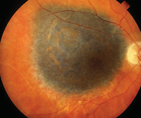
Choroidal melanoma is the most common primary intraocular malignancy in adults, and while it is a rare tumor overall, correctly identifying it — and distinguishing it from the far more common choroidal nevus — carries high stakes, because untreated growth threatens not only the eye but, through hematogenous spread most often to the liver, the patient’s life.
This site’s separate coverage of choroidal nevus versus small melanoma addresses the specific risk factors used to distinguish a suspicious nevus from an early melanoma; this article focuses on the melanoma itself once that distinction has been made.
Clinical Presentation
Many choroidal melanomas are discovered incidentally on a routine dilated fundus exam, before they have caused any visual symptoms, which is part of why regular eye exams have genuine value in catching this tumor early, sometimes before it has grown large enough to threaten either vision or systemic health.
When symptomatic, patients may report blurred vision, a visual field defect corresponding to the tumor’s location, photopsia, or, with more posterior or larger tumors, symptoms related to an associated exudative retinal detachment overlying the mass.
Exam Findings
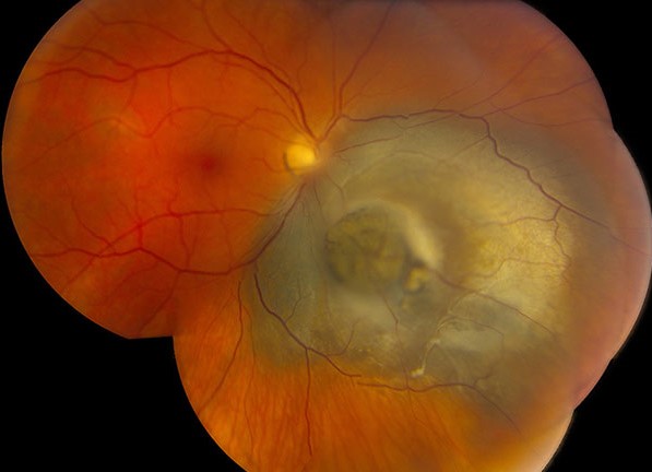
- A pigmented (or, in a meaningful minority of cases, amelanotic) elevated choroidal mass, classically dome-shaped, though larger tumors that have broken through Bruch’s membrane can take on a distinctive “mushroom” or “collar-button” configuration
- Orange pigment (lipofuscin) overlying the tumor surface — a feature specifically associated with more active, growing lesions and one of several risk factors used to distinguish melanoma from a stable nevus, as discussed in more detail in this site’s dedicated comparison article
- Subretinal fluid and, in some cases, a secondary exudative retinal detachment extending beyond the tumor margins
- Documented growth on serial exam or imaging — the single most decisive feature distinguishing an active melanoma from a stable, longstanding nevus, since nevi by definition remain stable over time
Fundus Explorer Pro
Photograph the retinal findings described here with the phone already in your pocket — 22 D optics and built-in illumination in one handheld unit.
From Choroida — the team behind this siteDiagnostic Evaluation
B-scan ultrasonography is central to the workup, characteristically showing a dome-shaped or mushroom-configured mass with low-to-medium internal reflectivity, a pattern that, together with other ultrasound features (choroidal excavation, orbital shadowing), helps distinguish melanoma from other choroidal masses, including the high-reflectivity pattern typical of choroidal hemangioma, discussed in its own dedicated article on this site.
Fundus photography and autofluorescence imaging document the tumor’s appearance and the presence of orange pigment, and serial photography over time is often what actually confirms growth when a single exam leaves genuine uncertainty about whether a pigmented lesion is a stable nevus or an early, growing melanoma.
Once melanoma is diagnosed or strongly suspected, systemic staging is essential given the tumor’s metastatic potential: liver function tests and liver imaging (given the liver’s status as the overwhelmingly predominant site of metastatic spread), along with genetic/cytogenetic testing of tumor tissue when biopsy is performed, since specific chromosomal and gene expression profiling findings (monosomy 3, among others) carry substantial prognostic value for predicting metastatic risk, independent of the tumor’s basic size and location.
Differential Diagnosis
- Choroidal nevus — the most important and most common alternative diagnosis, distinguished using the specific risk factors detailed in this site’s dedicated comparison article
- Choroidal metastasis — typically less elevated, sometimes multiple or bilateral, with a relevant known primary malignancy, discussed in its own dedicated article on this site
- Choroidal hemangioma — a distinctly different, high-reflectivity ultrasound pattern and orange-red rather than typically pigmented fundus appearance, discussed in its own dedicated article on this site
- Peripheral exudative hemorrhagic chorioretinopathy — a recognized melanoma mimic in older patients, discussed in its own dedicated article on this site, emphasizing how genuinely important careful multimodal evaluation is before committing to a melanoma diagnosis and its associated treatment
Management
Treatment is individualized based on tumor size, location, and the patient’s overall clinical picture, generally decided in consultation with an ocular oncology specialist familiar with the full range of available options.
Plaque brachytherapy (radioactive plaque applied to the sclera overlying the tumor) is the most commonly used eye-preserving treatment for small to medium tumors, having been shown in landmark trials to achieve survival outcomes statistically comparable to enucleation for appropriately selected tumors while preserving the eye and, often, useful vision.
Enucleation remains necessary for large tumors, tumors causing significant pain from secondary glaucoma, or eyes with no reasonable chance of preserving useful vision regardless of treatment approach, and the decision is made after weighing tumor characteristics against the realistic likelihood of achieving both local tumor control and meaningful visual function.
Other treatment modalities — including various forms of external beam or particle radiotherapy, and local resection in carefully selected cases — are used in specific clinical circumstances, generally at specialized ocular oncology centers given the complexity of both tumor management and the systemic surveillance that follows.
Regardless of the local eye treatment chosen, ongoing systemic surveillance (typically periodic liver imaging and liver function testing) continues indefinitely given the tumor’s capacity for late metastatic spread, sometimes occurring many years after apparently successful treatment of the primary eye tumor.
This lifelong surveillance requirement is an important point to communicate clearly at the time of diagnosis, since patients understandably tend to focus on the immediate eye treatment and may not otherwise anticipate that systemic monitoring will remain part of their care indefinitely.


Document what you see
Two smartphone imaging tools built for everyday clinic use — one for the slit lamp, one for the fundus.
From Choroida — the team behind this siteReferences
- Collaborative Ocular Melanoma Study Group. The COMS randomized trial of iodine 125 brachytherapy for choroidal melanoma. Archives of Ophthalmology.
- Shields CL, Shields JA. Ocular melanoma: relatively rare but requiring respect. Clinics in Dermatology.
- American Academy of Ophthalmology. Basic and Clinical Science Course, Section 4: Ophthalmic Pathology and Intraocular Tumors.
Test yourself
A few questions straight from this article.
-
Choroidal melanoma is best described as which of the following in adults?
Choroidal melanoma is the most common primary intraocular malignancy of adults, and although rare overall, untreated growth threatens both the eye and life through metastatic spread. -
Which event explains the mushroom or collar-button shape seen in larger choroidal melanomas?
Choroidal melanoma is classically dome-shaped, but once a larger tumour breaks through Bruch's membrane it takes on the distinctive mushroom or collar-button configuration. -
The orange pigment seen overlying the surface of a choroidal melanoma represents which material?
Orange pigment on the tumour surface is lipofuscin, and it is specifically associated with more active, growing lesions rather than stable ones. -
Which single feature most decisively separates an active choroidal melanoma from a stable choroidal nevus?
Documented growth is the single most decisive distinguishing feature, because a nevus by definition remains stable over time. -
On B-scan ultrasonography, choroidal melanoma characteristically shows which internal reflectivity?
B-scan typically shows a dome-shaped or mushroom-configured mass with low-to-medium internal reflectivity, often with choroidal excavation and orbital shadowing. -
Which features typically suggest choroidal metastasis rather than primary choroidal melanoma?
Choroidal metastases are typically less elevated and may be multiple or bilateral, usually in a patient with a relevant known primary malignancy. -
Which cytogenetic finding in choroidal melanoma tissue carries substantial prognostic value for metastatic risk?
Chromosomal and gene expression profiling findings such as monosomy 3 predict metastatic risk independently of the tumour's basic size and location. -
Which treatment is most commonly used to preserve the eye in small to medium choroidal melanoma?
A radioactive plaque applied to the sclera overlying the tumour is the commonest eye-preserving option, with landmark trials showing survival statistically comparable to enucleation in appropriately selected tumours. -
Enucleation remains necessary in choroidal melanoma in which of these situations?
Enucleation is reserved for large tumours, eyes with significant pain from secondary glaucoma, and eyes with no reasonable chance of useful vision whatever treatment is used. -
After local treatment of choroidal melanoma, which ongoing systemic surveillance is required?
Liver imaging and liver function testing continue indefinitely because metastatic spread can occur many years after apparently successful treatment of the eye tumour.

Writes and reviews the clinical articles on Educate. Every post is checked against peer-reviewed sources before it goes live.


