
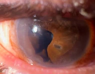
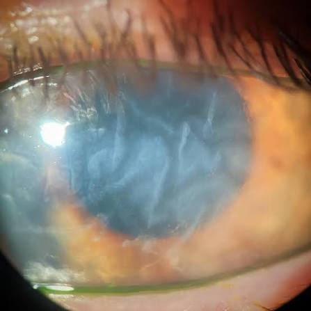
Descemet membrane folds and striae are wrinkles in the deepest, most posterior layer of the cornea, and because different underlying mechanisms produce folds with characteristically different orientations and distributions, learning to read the specific pattern gives a clinician real diagnostic information about what is stressing the cornea from behind, well before the underlying cause might otherwise become apparent.
Why Descemet Membrane Wrinkles
Descemet membrane is a thin, elastic basement membrane underlying the corneal endothelium, and it can develop visible folds or striae when subjected to mechanical compression, reduced intraocular pressure removing its normal supportive tension, or stromal swelling that distorts the posterior corneal architecture, each of which produces a somewhat different, recognizable pattern.
Hypotony-Related Folds
Low intraocular pressure, whether from surgical overfiltration, a wound leak, or other causes of hypotony, reduces the normal outward tension that helps keep Descemet membrane smooth, allowing folds to develop, typically with a somewhat irregular, often more diffuse or radial pattern corresponding to the generalized reduction in globe rigidity and corneal support.
- Recognizing hypotony-related folds should prompt assessment of intraocular pressure and, particularly in a recent postoperative patient, a careful search for a wound leak or other cause of reduced pressure
All-fit Slit-Lamp Adapter
Record and share exactly what you see at the slit lamp. One adapter fits any slit lamp or surgical microscope — and any smartphone.
From Choroida — the team behind this siteVertical Striae From Corneal Edema
When significant stromal edema develops, whether from endothelial dysfunction, acute pressure elevation, or other causes of corneal swelling, Descemet membrane can develop folds, classically described as vertical or near-vertical striae, as the swollen stroma distorts the underlying membrane, a pattern often seen together with the diffuse stromal haze characteristic of significant corneal edema (see corneal endothelial decompensation).
Postoperative and Intraoperative Folds
- Folds appearing in the immediate postoperative period after cataract or other intraocular surgery are common and often reflect a combination of transient surgically induced stromal edema and, in some cases, mechanical effects from the surgical wound or instrumentation
- Folds directly beneath or radiating from a surgical incision can reflect localized mechanical stress from wound construction or closure, generally resolving as the wound heals and stabilizes
- Most postoperative folds related to transient surgical trauma resolve spontaneously over days to weeks as associated edema clears and the eye stabilizes, distinguishing this generally self-limited pattern from folds related to an ongoing, unresolved underlying problem
Folds From External Compression
Folds can also develop from external mechanical compression of the globe, including from a tight bandage contact lens, eyelid pressure in conditions causing significant lid swelling, or other external sources of sustained pressure on the corneal surface, typically resolving once the compressive factor is identified and relieved.
Clinical Evaluation
- Careful slit-lamp assessment of the orientation, distribution, and depth of the folds, since this pattern recognition is central to narrowing the differential
- Intraocular pressure measurement, essential given the strong association between hypotony and one of the most common fold patterns
- A recent surgical history, and examination for any wound leak (including Seidel testing when appropriate) in a postoperative patient with new folds
- Assessment of overall corneal clarity and thickness, since folds accompanying significant diffuse edema point toward an endothelial or acute pressure-related process rather than a purely mechanical or hypotony-related cause
Management
Descemet membrane folds themselves are not directly treated, since management is entirely focused on identifying and correcting the underlying cause producing them.
- Hypotony-related folds are managed by identifying and addressing the cause of low pressure, whether a wound leak requiring surgical revision or another treatable cause of reduced aqueous production or outflow imbalance
- Folds related to transient postoperative edema generally require only observation, since they resolve as the eye heals
- Folds accompanying significant corneal edema from endothelial dysfunction are managed according to the underlying endothelial condition driving the edema
Clinical approach to folds
Descemet folds are a nonspecific sign of corneal stress. Ask about recent surgery, trauma, and inflammation, and measure the intraocular pressure, since both hypotony and high pressure can cause folds. Examine the stroma for edema and the endothelium for guttae and cells. If folds persist after a cataract operation, look for a Descemet detachment with OCT, which changes management. Observe if the cornea is clearing, and treat the cause if it is not.
Prognosis
Most Descemet membrane folds, once their underlying cause is identified and appropriately addressed, resolve over time without permanent consequence to corneal clarity or function.
Persistent, longstanding folds related to chronic hypotony or unresolved endothelial dysfunction carry more concern for lasting corneal changes, which is why prompt identification of the underlying driver, rather than simply observing the fold pattern itself, is the genuinely useful clinical habit this finding should prompt.


Document what you see
Two smartphone imaging tools built for everyday clinic use — one for the slit lamp, one for the fundus.
From Choroida — the team behind this siteReferences
- Waring GO 3rd, Bourne WM, Edelhauser HF, Kenyon KR. The corneal endothelium. Normal and pathologic structure and function. Ophthalmology. 1982;89:531-590.
- Costa VP, Spaeth GL, Eiferman RA, Orengo-Nania S. Wound healing modulation in glaucoma filtration surgery. Ophthalmic Surg. 1993;24:152-170.
- Fine IH, Hoffman RS. Phacoemulsification in the presence of pseudoexfoliation: challenges and options. J Cataract Refract Surg. 1997;23:160-165.
- Bourne WM. Biology of the corneal endothelium in health and disease. Eye (Lond). 2003;17:912-918.
