
Panretinal photocoagulation trades peripheral retina for central vision – deliberately, and with over fifty years of evidence behind the trade.
The Diabetic Retinopathy Study proved in the 1970s that scattering thousands of laser burns across the retinal periphery could cut severe vision loss from proliferative diabetic retinopathy by more than half.
Panretinal photocoagulation (PRP) remains, decades later, one of the most effective preventive treatments in all of ophthalmology.
Anti-VEGF therapy has now given clinicians a genuine alternative for some patients.
Knowing which patients still need the laser, and which can be managed with injections instead, is now a real clinical decision rather than a foregone conclusion.
What Is Panretinal Photocoagulation?
Panretinal photocoagulation is the application of thousands of laser burns across the peripheral retina, sparing the macula, to treat proliferative diabetic retinopathy and other ischaemic retinal diseases.
It is used across a range of conditions united by retinal ischaemia driving neovascularisation:
- Proliferative diabetic retinopathy – the original and still the commonest indication
- Ischaemic central and branch retinal vein occlusion with neovascularisation
- Neovascular glaucoma, to regress the ischaemic drive behind angle neovascularisation
- Retinopathy of prematurity, in a modified peripheral pattern in appropriate zones
- Sickle cell retinopathy with proliferative disease
What unites all of these is the same underlying mechanism – the laser interrupts a shared final common pathway, not just diabetic disease specifically.
Mechanism of Action
The logic of the treatment is counter-intuitive on first encounter: destroying tissue to preserve function.
- Ischaemic peripheral retina secretes VEGF and other angiogenic factors, driving abnormal new vessel growth at the disc and elsewhere
- Laser burns destroy the oxygen-hungry outer retinal photoreceptors across a large peripheral area
- Reduced peripheral oxygen demand allows the remaining choroidal circulation to adequately oxygenate the thinner residual retina
- This reduces the ischaemic drive for VEGF production, causing regression of neovascularisation over weeks to months
- The treatment does not restore any lost peripheral function – it converts ischaemic, VEGF-producing tissue into scarred, quiescent tissue
Explaining this trade-off clearly is essential, because patients understandably resist a treatment framed as “burning the retina” until they understand what it is protecting.
Fundus Explorer Pro
Photograph the retinal findings described here with the phone already in your pocket — 22 D optics and built-in illumination in one handheld unit.
From Choroida — the team behind this siteIndications and Evidence
- The Diabetic Retinopathy Study established that PRP reduces severe visual loss in proliferative diabetic retinopathy by more than half compared with no treatment
- The Early Treatment Diabetic Retinopathy Study refined timing, showing high-risk proliferative disease benefits from prompt treatment while very early proliferative disease may sometimes be observed
- High-risk characteristics guiding urgent treatment include neovascularisation of the disc of a defined extent, or any neovascularisation with vitreous or preretinal haemorrhage
- The DRCR Retina Network’s Protocol S demonstrated that anti-VEGF monotherapy achieves visual outcomes non-inferior to PRP in proliferative diabetic retinopathy over two years, with fewer visual field and peripheral vision side effects
Protocol S is the study that genuinely changed practice – it did not replace PRP, but it proved anti-VEGF was a legitimate primary alternative rather than just an adjunct.
Procedure
Planning
- Confirmation of proliferative disease and high-risk characteristics on dilated examination and, where available, ultra-widefield imaging
- Discussion of the anti-VEGF alternative where appropriate, factoring in the patient’s ability to attend frequent follow-up
Laser Application
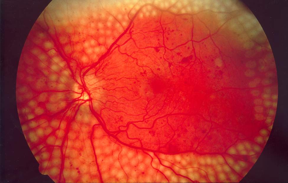
- Several thousand burns are placed across the mid and far periphery in one or more sessions, typically staged over two to four visits to reduce the risk of acute complications
- The macula, papillomacular bundle and major vascular arcades are deliberately spared
- Burn intensity is titrated to produce a moderate, whitish-grey retinal reaction without excessive intensity
- Full treatment typically requires 1200 to 1600 or more burns in total across sessions
Follow-Up
- Review at four to six weeks to assess regression of neovascularisation
- Additional “fill-in” laser if regression is incomplete
- Ongoing monitoring for recurrence, since regression does not guarantee the disease will not reactivate
Staging treatment across multiple sessions rather than a single mega-session is standard precisely to reduce the acute complications discussed below.
Complications
- Peripheral visual field constriction – expected and dose-related, sometimes affecting driving eligibility
- Reduced night vision and dark adaptation, from destruction of peripheral rod photoreceptors
- Transient or, less often, sustained macular oedema following treatment, particularly with aggressive single-session laser
- Choroidal effusion and transient angle-closure, more likely with heavy treatment delivered in one sitting
- Pain during treatment, usually manageable with topical anaesthesia and, when needed, sub-Tenon or peribulbar block
- Inadvertent foveal or vascular arcade burns from poor technique, causing permanent central field loss
Field constriction and impaired night vision are not complications in the strict sense – they are the expected cost of the treatment, and patients need to hear that distinction explicitly.
PRP Versus Anti-VEGF: Choosing Between Them
- Anti-VEGF avoids the peripheral field loss and night vision impairment of PRP, an advantage for younger, working patients
- PRP delivers a durable anatomical effect after a defined treatment course, without a requirement for indefinite injections
- Anti-VEGF requires sustained adherence to frequent injections; disease reactivates rapidly if treatment lapses, whereas PRP’s effect persists even if follow-up is interrupted
- Combination treatment – anti-VEGF to control acute disease activity with staged PRP for durability – is commonly used in practice, particularly where follow-up reliability is uncertain
The choice increasingly comes down to a single practical question: can this specific patient be relied upon to attend injections indefinitely? If not, PRP’s durability becomes the deciding factor.
Prognosis
PRP remains a highly effective treatment when applied at the right stage of disease.
- Adequately treated high-risk proliferative diabetic retinopathy has a substantially reduced risk of severe vision loss compared with untreated disease
- A minority of eyes fail to regress fully and require additional laser or adjunctive anti-VEGF therapy
- Visual field and night vision effects are permanent, and patients should be counselled about this before treatment, not after
- Long-term outcomes are best when PRP is combined with tight glycaemic and blood pressure control, since these remain the dominant drivers of overall diabetic eye disease progression
Fifty years on, PRP is still the treatment that reliably prevents blindness at scale – anti-VEGF has added options, not replaced the need to understand when the laser remains the right choice.


Document what you see
Two smartphone imaging tools built for everyday clinic use — one for the slit lamp, one for the fundus.
From Choroida — the team behind this siteReferences
- Diabetic Retinopathy Study Research Group. Photocoagulation treatment of proliferative diabetic retinopathy. Ophthalmology. 1981.
- Writing Committee for the DRCR Retina Network. Panretinal photocoagulation vs intravitreal ranibizumab for proliferative diabetic retinopathy (Protocol S). JAMA. 2015.
- Early Treatment Diabetic Retinopathy Study Research Group. Treatment techniques and clinical guidelines for photocoagulation (ETDRS Report 3). Ophthalmology. 1987.
- Panretinal Photocoagulation. EyeWiki, American Academy of Ophthalmology.
- Diabetic Retinopathy, Proliferative. StatPearls, NCBI Bookshelf.
Test yourself
A few questions straight from this article.
-
Which part of the retina is deliberately spared during panretinal photocoagulation?
PRP places thousands of burns across the mid and far periphery while deliberately sparing the macula, papillomacular bundle and major vascular arcades. -
How does panretinal photocoagulation reduce neovascularisation in ischaemic retinal disease?
Destroying oxygen-hungry peripheral photoreceptors reduces oxygen demand, letting the choroid oxygenate the residual retina and cutting the ischaemic drive for VEGF. -
Roughly how many laser burns does a full course of panretinal photocoagulation typically require?
Full treatment typically requires 1200 to 1600 or more burns in total, usually staged across two to four sessions. -
What did the Diabetic Retinopathy Study establish about panretinal photocoagulation in proliferative diabetic retinopathy?
The Diabetic Retinopathy Study showed that scatter laser cut severe visual loss from proliferative diabetic retinopathy by more than half compared with no treatment. -
What did Protocol S show about anti-VEGF monotherapy in proliferative diabetic retinopathy?
The DRCR Retina Network's Protocol S found anti-VEGF monotherapy non-inferior to PRP over two years, with fewer visual field and peripheral vision side effects. -
Which finding counts as a high-risk characteristic demanding prompt panretinal photocoagulation?
High-risk characteristics include neovascularisation of the disc of a defined extent, or any neovascularisation accompanied by vitreous or preretinal haemorrhage. -
Why is panretinal photocoagulation usually staged across two to four sessions rather than delivered in one sitting?
Staging treatment reduces the risk of acute complications, including post-treatment macular oedema, choroidal effusion and transient angle closure seen with heavy single-session laser. -
Which effect of panretinal photocoagulation should be presented to patients as an expected cost rather than a complication?
Field constriction and impaired dark adaptation follow inevitably from destroying peripheral rods; the article stresses patients must hear this distinction explicitly before treatment. -
Which practical question most often decides between PRP and anti-VEGF for proliferative diabetic retinopathy?
Anti-VEGF disease reactivates rapidly if treatment lapses, whereas PRP's effect persists through interrupted follow-up, so reliability of attendance becomes the deciding factor. -
What most improves long-term outcomes in eyes treated with panretinal photocoagulation?
Long-term outcomes are best when PRP is combined with tight glycaemic and blood pressure control, which remain the dominant drivers of diabetic eye disease progression.

Writes and reviews the clinical articles on Educate. Every post is checked against peer-reviewed sources before it goes live.


