
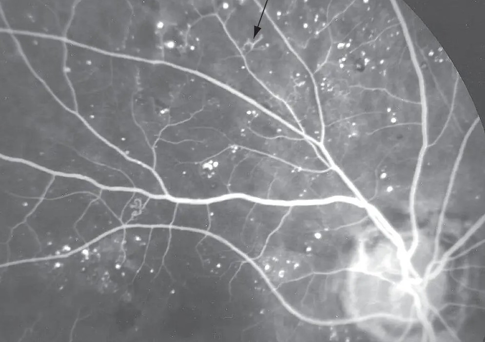
Intraretinal microvascular abnormalities (IRMA) are shunt vessels and new intraretinal vessels that develop in areas of capillary nonperfusion in diabetic retinopathy.
They are also a sign of severe ischemia, and they are one of the three features of severe nonproliferative retinopathy.
The practical difficulty is that they can look like early neovascularization, and the distinction affects treatment.
What Are Intraretinal Microvascular Abnormalities?
IRMA are tortuous, dilated capillary segments that lie within the retina, adjacent to areas of capillary closure.
They are thought to be either remodeled preexisting capillaries or intraretinal new vessels that have not yet broken through the internal limiting membrane.
Because they remain within the retina, they do not leak into the vitreous and do not cause hemorrhage in the same way as neovascularization.
They appear when retinal ischemia reaches a degree that stimulates vascular endothelial growth factor.
The ETDRS found that IRMA, venous beading, and extensive hemorrhages predicted progression to proliferative disease.
Clinical Appearance
- Fine, irregular, red intraretinal lines that run between arterioles and venules, usually at the border of nonperfused retina
- Located in the posterior pole or the midperiphery, often near areas of cotton wool spots or hemorrhages
- Without crossing major retinal vessels and without protruding into the vitreous
- Often accompanied by venous beading and loops
- Best seen with a red-free filter or a high magnification contact lens
They look like faint tangles and are sometimes mistaken for background microaneurysms.
Fundus Explorer Pro
Photograph the retinal findings described here with the phone already in your pocket — 22 D optics and built-in illumination in one handheld unit.
From Choroida — the team behind this siteHow to Distinguish IRMA From Neovascularization
Retinal neovascularization grows on the surface of the retina or the disc, forms flat networks or fronds, and crosses large vessels.
In addition, IRMA stays in the plane of the retina and does not cross large vessels.
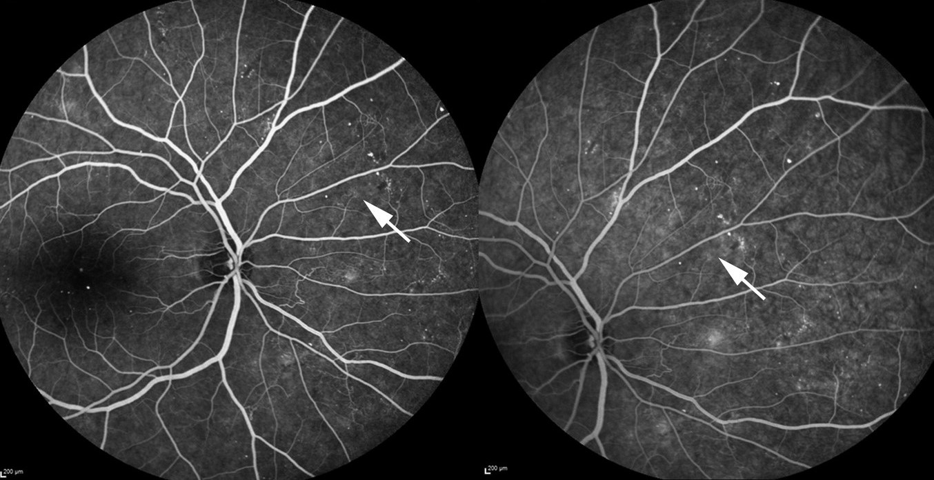
On fluorescein angiography, neovascularization leaks profusely and early, while IRMA shows mild leakage or none and stays within the retinal layers (see fluorescein angiography patterns).
OCT angiography can also show IRMA as abnormal vessels in the inner retinal layers and neovascularization as flow above the internal limiting membrane (see OCT angiography).
When in doubt, angiography is useful, since treating a patient as if neovascularization is present changes both the urgency and the follow-up.
Clinical Significance
- IRMA indicates widespread ischemia and an increased risk of progression to proliferative disease within a year
- Moderate to severe IRMA is one component of the 4-2-1 rule (see diabetic retinopathy classification)
- Clinicians may see IRMA with other ischemic conditions, including retinal vein occlusion and ocular ischemic syndrome
- It can regress after panretinal photocoagulation or anti-VEGF therapy
Patients with IRMA need closer follow-up, with examination intervals of weeks to a few months, according to the other findings and the guideline.
Management for Intraretinal Microvascular Abnormalities
Management follows the stage of retinopathy and the presence of macular edema.
Eyes with severe nonproliferative disease may be observed closely, or treated early with panretinal photocoagulation or anti-VEGF injections, especially when follow-up is unreliable (see panretinal photocoagulation and anti-VEGF therapy).
Control of blood glucose, blood pressure, and lipids is essential.
A rapid improvement in glycemic control can briefly worsen retinopathy.
Pregnant patients and those scheduled for cataract surgery need particular attention, because progression can be rapid (see diabetic retinopathy in pregnancy).
Tell patients that the finding is a warning sign and not yet a threat to vision, and that regular follow-up is the way to catch the next step.
A Short Practical Checklist
- Examine with a red-free filter and a high-power lens, and look at the border of nonperfused retina
- Compare the vessel with a neighboring venule: IRMA is finer and more irregular
- Ask whether the vessel crosses a major vein or arteriole, which suggests neovascularization
- Obtain angiography when the result will change the decision
- Look for IRMA in the periphery with a widefield image
Intraretinal Microvascular Abnormalities: Treatment Timing
Studies of early panretinal photocoagulation in severe nonproliferative disease showed a reduction in progression, with a modest absolute benefit. Anti-VEGF agents, in contrast, can reverse the severity level without necessarily changing the underlying ischemia.
Because stopping anti-VEGF treatment can lead to a return of disease, patients who start it need to be committed to follow-up.
The choice is individualized according to the patient’s reliability, the other eye, and the presence of macular edema.
Document IRMA with a drawing or a photograph at the first visit.
Patients who are pregnant, have rapidly improved their glucose levels, or are due for surgery on the eye deserve a shorter interval to the next examination.
For the latest studies, see the PubMed results for intraretinal microvascular abnormalities.


Document what you see
Two smartphone imaging tools built for everyday clinic use — one for the slit lamp, one for the fundus.
From Choroida — the team behind this siteReferences
- Early Treatment Diabetic Retinopathy Study Research Group. Fundus photographic risk factors for progression of diabetic retinopathy. ETDRS report number 12. Ophthalmology. 1991;98:823-833.
- Wilkinson CP, Ferris FL III, Klein RE, et al. Proposed international clinical diabetic retinopathy and diabetic macular edema disease severity scales. Ophthalmology. 2003;110:1677-1682.
- Ryan SJ, Schachat AP, Wilkinson CP, et al, eds. Retina. 6th ed. Elsevier; 2018.
- American Academy of Ophthalmology. Basic and Clinical Science Course, Section 12: Retina and Vitreous. AAO; current edition.

Writes and reviews the clinical articles on Educate. Every post is checked against peer-reviewed sources before it goes live.


