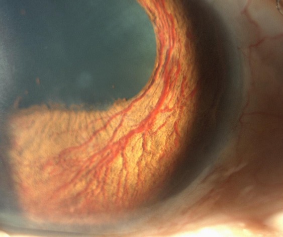
Rubeosis iridis — new blood vessel growth on the anterior surface of the iris — is the visible precursor to neovascular glaucoma.
Recognizing it before it progresses to angle involvement and secondary glaucoma is one of the more time-sensitive tasks in retinal and glaucoma care, since the disease can move from an entirely reversible early stage to permanent, sight-threatening angle scarring in a matter of weeks.
The finding itself is a direct consequence of retinal ischemia, which drives release of vascular endothelial growth factor (VEGF) into the aqueous, where it diffuses forward and stimulates abnormal vessel growth on iris tissue that is not normally vascularized this way.
Pathogenesis
Any condition producing significant retinal ischemia can elevate intraocular VEGF enough to drive rubeosis, which is why the finding functions as a shared final pathway across several unrelated retinal diseases rather than being specific to any one of them.
The new vessels themselves are fragile, lack normal tight junctions, and are prone to leaking and bleeding, which contributes both to the clinical findings (fine, irregular vessels crossing the pupillary margin) and to the eventual fibrovascular membrane that contracts across the angle to cause neovascular glaucoma.
This same VEGF-driven mechanism underlies neovascularization elsewhere in the eye — the retina and optic disc in proliferative diabetic retinopathy, for example — which is why rubeosis so often coexists with other neovascular complications in the same ischemic eye rather than occurring as an isolated finding.
Underlying Causes
- Proliferative diabetic retinopathy — one of the most common causes overall
- Central retinal vein occlusion, particularly the ischemic form — historically associated with such a high rate of neovascular glaucoma that the complication was once called “90-day glaucoma” for its typical timing after the occlusion
- Ocular ischemic syndrome, from severe carotid artery stenosis or occlusion
- Chronic retinal detachment
- Central retinal artery occlusion, less commonly than vein occlusion but still a recognized cause
- Intraocular tumors and chronic uveitis, less common causes
All-fit Slit-Lamp Adapter
Record and share exactly what you see at the slit lamp. One adapter fits any slit lamp or surgical microscope — and any smartphone.
From Choroida — the team behind this siteClinical Findings and Staging
- Earliest sign: fine, irregular, tuft-like vessels at the pupillary margin, easily missed without a careful, undilated slit-lamp exam of the iris (dilation can mask or alter the appearance of early vessels)
- Progression to vessels crossing the iris surface toward the angle
- Gonioscopic evidence of angle neovascularization, a critical finding that precedes or accompanies pressure elevation
- Peripheral anterior synechiae as the fibrovascular membrane contracts, progressively closing the angle
- Elevated intraocular pressure once angle involvement is significant, marking the transition to frank neovascular glaucoma
The exam should specifically include careful inspection of the pupillary margin before dilation, because that is where the earliest, most subtle vessels are found.
Gonioscopy is essential whenever rubeosis is suspected or a known ischemic retinal condition is being monitored, because angle findings often precede any visible pressure elevation, and a normal intraocular pressure reading should never be used to defer gonioscopy in a patient with a known high-risk ischemic condition.
Differential Diagnosis
- Normal iris vasculature — radial iris vessels are a normal anatomic finding and should not be mistaken for rubeosis; the key distinguishing feature of rubeosis is fine, disorganized, leaky-appearing new vessels rather than the normal radial pattern
- Iris nevus or melanoma with associated vascularity — a mass lesion is usually apparent alongside any vascular change
- Chronic anterior uveitis with iris hyperemia — diffuse injection rather than discrete abnormal vessels, with other signs of active inflammation
Management
The priority once rubeosis is identified is treating the underlying retinal ischemia driving VEGF production.
Panretinal photocoagulation remains the standard treatment for ischemic retinopathy from diabetes, vein occlusion, or other causes, and can induce regression of iris neovascularization when applied promptly, before angle changes become fixed and permanent, by reducing the ischemic retinal tissue driving ongoing VEGF production.
Intravitreal anti-VEGF injections produce rapid, though temporary, regression of the new vessels and are frequently used as an adjunct — either to gain quick control while panretinal photocoagulation is being completed, or preoperatively to reduce intraoperative bleeding risk if surgery is needed.
Because the effect of anti-VEGF injection is temporary and the underlying ischemic drive persists unless definitively treated, repeated injections without also completing panretinal photocoagulation or otherwise addressing the ischemia tend to be a temporizing measure rather than a durable solution on their own.
Once angle involvement and elevated pressure develop, management shifts toward neovascular glaucoma itself: medical therapy to lower pressure, and, when medical therapy is insufficient, glaucoma drainage device surgery or cyclophotocoagulation, because standard trabeculectomy tends to fail at a high rate with active neovascularization and its associated scarring, given the tendency of new vessels to bleed into and obstruct a conventional filtering bleb.
Why Early Recognition Matters
Once peripheral anterior synechiae have formed and the angle is significantly compromised, the process is irreversible even with successful treatment of the underlying ischemia.
Panretinal photocoagulation and anti-VEGF therapy can regress the visible new vessels, but they cannot open an angle that has already scarred closed.
This is the central argument for regular, careful anterior segment and gonioscopic surveillance in any patient with a known retinal ischemic condition, well before pressure elevation or symptoms develop, since the window for genuinely preventing neovascular glaucoma closes well before the disease becomes clinically obvious.


Document what you see
Two smartphone imaging tools built for everyday clinic use — one for the slit lamp, one for the fundus.
From Choroida — the team behind this siteReferences
- Sivak-Callcott JA, O’Day DM, Gass JD, Tsai JC. Evidence-based recommendations for the diagnosis and treatment of neovascular glaucoma. Ophthalmology.
- Hayreh SS. Neovascular glaucoma. Progress in Retinal and Eye Research.
- American Academy of Ophthalmology. Basic and Clinical Science Course, Section 10: Glaucoma.
- Rodrigues GB, Abe RY, Zangalli C, et al. Neovascular glaucoma: a review. International Journal of Retina and Vitreous.
Test yourself
A few questions straight from this article.
-
Rubeosis iridis refers to which clinical finding?
Rubeosis iridis is new blood vessel growth on the anterior surface of the iris, and is the visible precursor to neovascular glaucoma. -
What drives the development of rubeosis iridis?
Retinal ischaemia releases vascular endothelial growth factor into the aqueous, which diffuses forward and stimulates abnormal vessel growth on the iris. -
Where is the earliest sign of rubeosis iridis found on slit-lamp examination?
The earliest vessels are fine, irregular and tuft-like at the pupillary margin, and are easily missed without a careful undilated slit-lamp exam. -
Which retinal disease is among the most common causes of rubeosis iridis overall?
Proliferative diabetic retinopathy is one of the most common causes overall, alongside ischaemic vein occlusion and ocular ischaemic syndrome. -
A patient with ischaemic retinopathy has suspected rubeosis iridis and a normal intraocular pressure. What is the next step?
Angle neovascularisation often precedes any pressure elevation, so a normal intraocular pressure should never be used to defer gonioscopy. -
Which feature distinguishes rubeosis iridis from normal iris vasculature?
Radial iris vessels are a normal anatomic finding; rubeosis is distinguished by fine, disorganised, leaky-appearing new vessels. -
What is the standard treatment for the retinal ischaemia driving rubeosis iridis?
Panretinal photocoagulation reduces the ischaemic retinal tissue driving VEGF production and can regress iris neovascularisation if applied promptly. -
What is the main limitation of intravitreal anti-VEGF injection in rubeosis iridis?
Anti-VEGF produces rapid but temporary regression, so repeated injections without treating the ischaemia are only a temporising measure. -
Which surgical option is preferred in neovascular glaucoma when medical pressure-lowering therapy is insufficient?
Standard trabeculectomy fails at a high rate with active neovascularisation, so a glaucoma drainage device or cyclophotocoagulation is preferred. -
Once peripheral anterior synechiae have significantly closed the angle in neovascular glaucoma, what is the outcome?
Treatment can regress visible new vessels but cannot open an angle that has already scarred closed, which is why early surveillance matters.

Writes and reviews the clinical articles on Educate. Every post is checked against peer-reviewed sources before it goes live.