
Proliferative diabetic retinopathy (PDR) is the stage of diabetic retinopathy in which new vessels grow on the optic disc, elsewhere on the retina, or on the iris.
These vessels bleed, contract, and pull on the retina, and they are the main route to severe vision loss in diabetes.
Panretinal photocoagulation (PRP) was the standard treatment for decades, and anti-VEGF injections have since become a real alternative.
The choice between them often turns on whether the patient can return for follow-up.
Both approaches lower the risk of severe vision loss, and each has drawbacks that matter differently from one patient to the next.
This article reviews the clinical course of PDR, what each treatment achieves, where the evidence stands, and when vitrectomy becomes necessary.
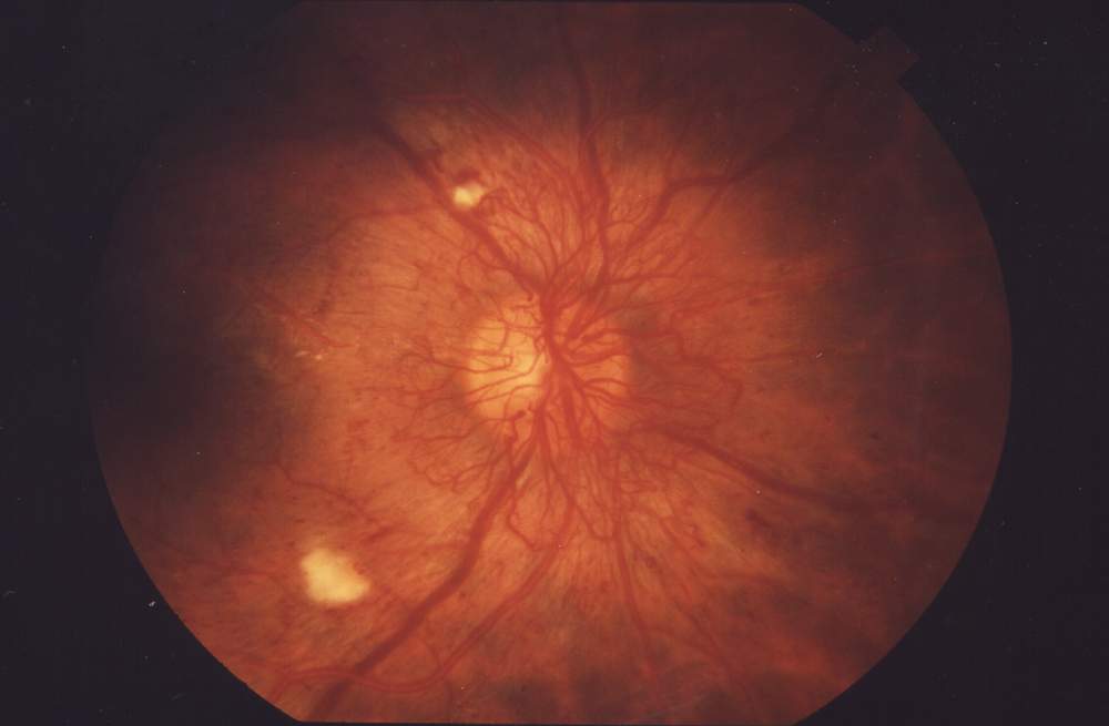
What Defines Proliferative Diabetic Retinopathy?
PDR is defined by neovascularization of the disc (NVD) or elsewhere (NVE), and by its complications: preretinal hemorrhage, vitreous hemorrhage, and fibrovascular proliferation.
Retinal ischemia drives VEGF release, and VEGF drives new vessel growth.
The new vessels lack normal barrier function and grow along the posterior hyaloid, where vitreous traction tears them.
The Diabetic Retinopathy Study defined high-risk characteristics that carry a high chance of severe vision loss without treatment:
- NVD of one-quarter to one-third of a disc area or more, with or without vitreous hemorrhage
- Any NVD with vitreous or preretinal hemorrhage
- NVE of at least half a disc area with vitreous or preretinal hemorrhage
Clinical Course
Many patients have no symptoms until a vitreous hemorrhage causes floaters or sudden blur.
The neovascular fronds themselves are found on dilated examination and confirmed with widefield fluorescein angiography, which also maps capillary nonperfusion.
Fibrovascular tissue may contract over months and cause tractional retinal detachment, which threatens the macula (see tractional retinal detachment).
Rubeosis iridis and neovascular glaucoma follow when ischemia is severe (see neovascular glaucoma).
A hemorrhage that obscures the fundus does not lower the need for careful evaluation of the rest of the retina and of the fellow eye (see vitreous hemorrhage).
Fundus Explorer Pro
Photograph the retinal findings described here with the phone already in your pocket — 22 D optics and built-in illumination in one handheld unit.
From Choroida — the team behind this siteDiagnosis and Imaging
Neovascularization on the disc can be subtle, and a fine loop of vessels lying flat on the disc surface is easy to miss.
Fluorescein angiography shows early, intense, and progressive leakage from the new vessels, unlike normal disc vessels, which do not leak.
Widefield angiography is particularly useful because a large share of diabetic lesions and nonperfusion lies outside the standard seven fields.
OCT helps identify vitreoretinal traction, macular edema, and the extent of fibrovascular tissue, and OCT angiography can show neovascular fronds without dye.
The iris and angle should be examined before dilation in every eye with severe ischemia, because early rubeosis is easy to miss once the pupil is wide.
Panretinal Photocoagulation
PRP destroys ischemic peripheral retina, lowers oxygen demand, and reduces the VEGF stimulus.
The Diabetic Retinopathy Study showed that PRP reduced the risk of severe vision loss by more than half in eyes with high-risk PDR.
A typical session uses several hundred burns, and the full treatment often takes 1200 to 1600 or more burns of about 500 µm spread over more than one visit (see panretinal photocoagulation).
The drawbacks are real:
- Constriction of the peripheral visual field
- Reduced night vision and dark adaptation
- Worsening of macular edema, particularly with heavy single-session treatment
- Pain and, rarely, transient angle closure or choroidal effusion
PRP is permanent, requires no injections, and is reliable in patients who may not return.
Anti-VEGF Therapy
Anti-VEGF agents cause rapid regression of new vessels, and they treat coexisting DME at the same time.
DRCR Protocol S compared ranibizumab with PRP in PDR.
Ranibizumab was noninferior to PRP at two years, caused less visual field loss, and led to fewer vitrectomies and less development of DME.
The CLARITY trial found aflibercept superior to PRP for visual acuity at 52 weeks.
Longer follow-up in Protocol S showed similar acuity outcomes between the two approaches, but the regression of neovascularization with injections lasts only as long as the injections continue.
Eyes lost to follow-up after anti-VEGF alone have been reported to do worse than eyes that received PRP, and some develop advanced fibrovascular disease with tractional detachment.
The Crunch Phenomenon
Anti-VEGF injection can cause rapid contraction of fibrovascular tissue in eyes with a large fibrous component, a process sometimes called the crunch phenomenon.
Tractional retinal detachment developed within days to weeks in reported series (see crunch syndrome).
Eyes with established traction should be monitored closely after injection or treated with vitrectomy instead.
Choosing a Treatment
There is no single correct answer, and the choice depends as much on the patient as on the retina.
Factors that favor PRP:
- Poor or uncertain adherence to follow-up
- Inability to afford or access repeated injections
- Pregnancy, where anti-VEGF use is generally avoided
- A limited view that prevents a full assessment of the peripheral retina
Factors that favor anti-VEGF:
- Concurrent center-involving DME
- Visual field preservation is important to the patient
- Reliable attendance
- Cataract surgery planned, since PRP can worsen edema
Many retina specialists combine the two, using anti-VEGF for fast control and PRP for durable protection.
Vitrectomy
Pars plana vitrectomy is indicated for:
- Nonclearing vitreous hemorrhage
- Tractional retinal detachment involving or threatening the macula
- Combined tractional and rhegmatogenous detachment
- Dense premacular hemorrhage
- Progressive fibrovascular proliferation despite treatment
Some surgeons inject bevacizumab a few days before surgery to reduce intraoperative bleeding, and the delay should be short to avoid worsening traction (see pars plana vitrectomy).
Follow-Up and Prognosis
After treatment, eyes need review every few weeks at first, then at longer intervals once the vessels have regressed.
Fibrovascular tissue may persist as a whitish scar even when active leakage is gone.
Visual outcome depends mainly on macular status, the presence of traction, and how promptly PDR is treated.
Systemic control of glucose, blood pressure, and lipids remains part of every visit.


Document what you see
Two smartphone imaging tools built for everyday clinic use — one for the slit lamp, one for the fundus.
From Choroida — the team behind this siteReferences
- Diabetic Retinopathy Study Research Group. Photocoagulation treatment of proliferative diabetic retinopathy: clinical application of Diabetic Retinopathy Study findings. DRS report number 8. Ophthalmology. 1981;88:583-600.
- Gross JG, Glassman AR, Jampol LM, et al. Panretinal photocoagulation vs intravitreous ranibizumab for proliferative diabetic retinopathy: a randomized clinical trial. JAMA. 2015;314:2137-2146.
- Sivaprasad S, Prevost AT, Vasconcelos JC, et al. Clinical efficacy of intravitreal aflibercept versus panretinal photocoagulation for best corrected visual acuity in patients with proliferative diabetic retinopathy at 52 weeks (CLARITY): a multicentre, single-blinded, randomised, controlled, phase 2b, non-inferiority trial. Lancet. 2017;389:2193-2203.
- Obeid A, Su D, Patel SN, et al. Outcomes of eyes lost to follow-up with proliferative diabetic retinopathy that received panretinal photocoagulation versus intravitreal anti-vascular endothelial growth factor. Ophthalmology. 2019;126:407-413.
- Arevalo JF, Maia M, Flynn HW Jr, et al. Tractional retinal detachment following intravitreal bevacizumab (Avastin) in patients with severe proliferative diabetic retinopathy. Br J Ophthalmol. 2008;92:213-216.
Test yourself
A few questions straight from this article.
-
Proliferative diabetic retinopathy is defined by which finding?
PDR is the stage at which new vessels grow on the disc, elsewhere on the retina, or on the iris, together with complications such as preretinal and vitreous haemorrhage. -
What drives new vessel growth in proliferative diabetic retinopathy?
Ischaemic retina releases VEGF, which drives new vessel growth; these vessels lack normal barrier function and grow along the posterior hyaloid where traction tears them. -
Which extent of NVE with vitreous or preretinal haemorrhage counts as a high-risk characteristic in PDR?
The Diabetic Retinopathy Study listed NVE of at least half a disc area accompanied by vitreous or preretinal haemorrhage among the high-risk characteristics. -
In the Diabetic Retinopathy Study, how much did PRP reduce severe vision loss in high-risk PDR?
PRP destroys ischaemic peripheral retina and lowers the VEGF stimulus, and the Diabetic Retinopathy Study showed it cut the risk of severe vision loss by more than half. -
What did DRCR Protocol S show for ranibizumab compared with PRP in PDR at two years?
Protocol S found ranibizumab noninferior to PRP at two years, with less visual field loss, fewer vitrectomies and less development of diabetic macular oedema. -
What is the crunch phenomenon after anti-VEGF injection in proliferative diabetic retinopathy?
In eyes with a large fibrous component, injection can make fibrovascular tissue contract quickly, with tractional detachment reported within days to weeks. -
Which patient factor favours panretinal photocoagulation over anti-VEGF in PDR?
PRP is permanent and needs no injections, so it is the safer choice when a patient may not return; eyes lost to follow-up after anti-VEGF alone have done worse. -
Which finding favours anti-VEGF therapy rather than PRP in proliferative diabetic retinopathy?
Anti-VEGF regresses new vessels rapidly and treats coexisting macular oedema at the same time, whereas PRP can worsen that oedema. -
Which situation is an indication for pars plana vitrectomy in proliferative diabetic retinopathy?
Vitrectomy is indicated for non-clearing vitreous haemorrhage, tractional detachment involving or threatening the macula, dense premacular haemorrhage and progressive proliferation. -
On fluorescein angiography, what distinguishes disc neovascularisation from normal disc vessels?
New vessels leak early, intensely and progressively on angiography, while normal disc vessels do not leak at all.

Writes and reviews the clinical articles on Educate. Every post is checked against peer-reviewed sources before it goes live.


