
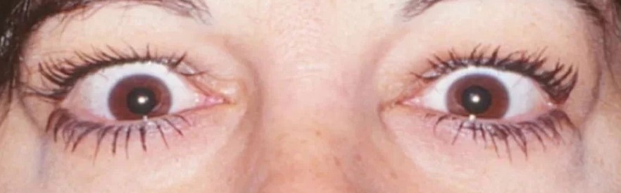
Eyelid retraction is the abnormal elevation of the upper eyelid margin or the depression of the lower eyelid margin, which exposes a rim of sclera.
Thyroid eye disease is the commonest cause, but it is not the only one, and a systematic approach finds the correct diagnosis.
The cause determines whether the patient needs imaging, endocrine tests, or surgery.
What Is Normal and What Is Eyelid Retraction?
The upper lid margin normally rests about one to two millimeters below the superior limbus, and the lower lid rests at or slightly above the inferior limbus.
Retraction is present when the upper lid margin lies above the limbus, exposing superior sclera, or when the lower lid sits below the limbus, exposing inferior sclera.
Measure the margin reflex distance, the width of the palpebral fissure, and the lid position on downgaze, to quantify the retraction and to detect lid lag.
Compare the two sides, because the apparent retraction of one lid may be a result of ptosis on the opposite side, as described by Hering’s law.
Causes
- Thyroid eye disease: the most frequent cause, from overaction of Müller muscle and fibrosis of the levator (see thyroid eye disease)
- Neurogenic: dorsal midbrain lesions with the Collier sign, aberrant regeneration of the oculomotor nerve, and Marcus Gunn jaw-winking (see Marcus Gunn jaw-winking syndrome)
- Mechanical or cicatricial: scarring of the eyelid after trauma, burns, or surgery, including overcorrection of ptosis or blepharoplasty (see upper eyelid blepharoplasty)
- Proptosis and high myopia: a large or prominent globe pushes the lid margins away (see proptosis differential diagnosis)
- Pseudoretraction: contralateral ptosis, or a facial nerve palsy with lower lid retraction
- Drugs and systemic causes: sympathomimetic agents, hyperthyroidism, and some neurologic disorders
- Congenital and idiopathic: a persistent, usually mild retraction from birth
All-fit Slit-Lamp Adapter
Record and share exactly what you see at the slit lamp. One adapter fits any slit lamp or surgical microscope — and any smartphone.
From Choroida — the team behind this siteEvaluation
Take a history that includes the onset, the course, symptoms of thyroid disease, trauma or surgery, and medications.
Measure the lid position in primary gaze and on downgaze, and look for lagophthalmos and corneal exposure.
Examine the pupils, the eye movements, and the globe position, since proptosis and ocular motility restriction suggest thyroid disease and an abnormal pupil or gaze suggests a brainstem disorder.
Check thyroid function and thyroid antibodies in adults without an obvious cause.
Perform imaging of the orbit when proptosis or a mass is suspected, and of the brain when neurological signs are present.
A phenylephrine test helps to distinguish pseudoretraction from true retraction, and to predict the effect of treatment.
Treatment
Treat the cause first, including control of thyroid function and the active phase of the thyroid eye disease.
Lubricants and ointment protect the cornea from exposure while the cause is being addressed (see exposure keratopathy).
Botulinum toxin or triamcinolone injection into the levator or Müller muscle can reduce retraction temporarily.
Surgery is done when the condition is stable, and the choice depends on the amount of retraction: recession of Müller muscle, levator recession, or a spacer graft for the lower lid.
Patients should know that surgery improves appearance and corneal protection, and that adjustments may be needed.
Clinical Clues to the Cause
Retraction that is bilateral, with proptosis, lid lag, and a history of thyroid disease, points to Graves orbitopathy, which is the commonest cause by far.
A retraction that is unilateral, with a ptotic lid on the other side, suggests Hering’s law at work, and covering the ptotic lid or lifting it manually will reveal that the retracted lid returns to a normal position.
Retraction on attempted upgaze with a fixed pupil and a poor convergence suggests a midbrain lesion, and the finding of retraction in a child with a swelling head and a setting-sun sign suggests hydrocephalus.
Retraction that follows eyelid, orbital, or facial surgery or trauma indicates scarring of the posterior or anterior lamella.
Special Situations
- Sympathetic drugs: apraclonidine and phenylephrine drops cause temporary retraction, which resolves when they are stopped
- Facial palsy: loss of orbicularis tone causes lower lid retraction and exposure
- Infants: transient retraction in newborns may be seen with prematurity or with raised intracranial pressure
- Cirrhosis and other systemic diseases: rare associations that are reported
Protecting the Eye
Whatever the cause, the cornea needs protection while the condition is addressed.
Use lubricant drops during the day and ointment at night, and consider a moisture chamber or taping the lids at night if lagophthalmos is present.
Look for signs of exposure keratopathy on fluorescein staining, and manage them early.
Counselling
Explain that the lids may return to a normal position when the cause is treated, and that surgery is a later option after stabilization.
Reassure patients that many causes are benign, but that new lid retraction in an adult deserves evaluation.


Document what you see
Two smartphone imaging tools built for everyday clinic use — one for the slit lamp, one for the fundus.
From Choroida — the team behind this siteReferences
- Bartley GB. The differential diagnosis and classification of eyelid retraction. Ophthalmology. 1996;103:168-176.
- Salmon JF. Kanski’s Clinical Ophthalmology: A Systematic Approach. 9th ed. Elsevier; 2020.
- Liu GT, Volpe NJ, Galetta SL. Neuro-Ophthalmology: Diagnosis and Management. 3rd ed. Elsevier; 2019.
- American Academy of Ophthalmology. Basic and Clinical Science Course, Section 7: Oculofacial Plastic and Orbital Surgery. AAO; current edition.

Writes and reviews the clinical articles on Educate. Every post is checked against peer-reviewed sources before it goes live.


