
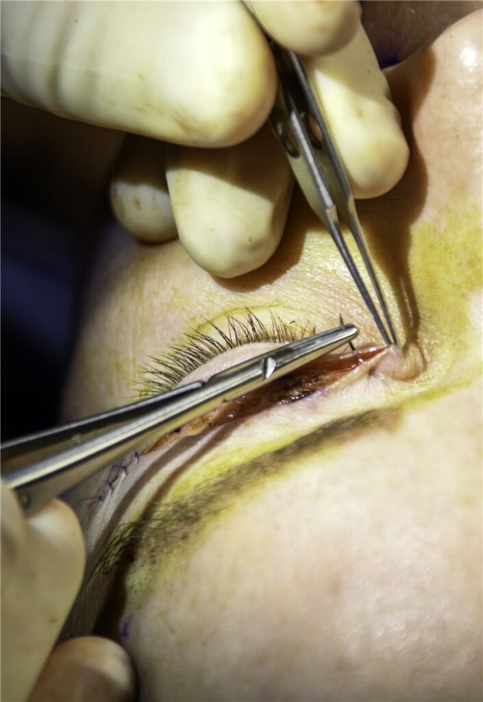
Upper eyelid blepharoplasty removes redundant skin, and often orbital fat, from the upper eyelid to correct dermatochalasis, the age-related laxity and excess skin that can hood the lid margin and, when severe enough, obstruct the superior visual field.
It sits at the boundary between cosmetic and functional surgery, and that boundary is exactly what determines whether a given case is approached as an elective aesthetic procedure or one with a genuine functional indication supported by visual field testing.
Dermatochalasis: The Underlying Problem
Skin laxity increases with age as collagen and elastin content in the eyelid skin decline, and this is compounded in the upper lid by weakening of the orbital septum, which allows orbital fat to prolapse forward and add to the visible bulk.
The result is redundant, often hooded skin overhanging the lid margin, sometimes accompanied by visible fat bulging, most commonly in the medial and central compartments of the upper lid.
When this excess skin is significant enough to fold over and mechanically obstruct the superior visual field, dermatochalasis moves from a cosmetic concern to a functional one, and this distinction is what the preoperative evaluation is really trying to establish.
Preoperative Evaluation
- Visual field testing, with and without the lid skin manually elevated (taped), to document the degree of field loss attributable to the skin itself and confirm it improves when the obstruction is lifted
- Assessment of eyelid position and function, including margin reflex distance and levator function, to distinguish dermatochalasis from coexisting ptosis, which is common in the same age group and needs to be addressed separately if present
- Examination for dry eye disease and adequate tear film, since blepharoplasty can worsen pre-existing dry eye by altering lid closure dynamics, at least temporarily
- Assessment of brow position, since a low or ptotic brow can mimic or worsen the appearance of dermatochalasis and may need to be addressed concurrently or instead of eyelid skin excision alone
Distinguishing true dermatochalasis from brow ptosis is a common pitfall: operating on the eyelid skin alone in a patient whose main problem is a descended brow gives an incomplete, sometimes unsatisfying result.
All-fit Slit-Lamp Adapter
Record and share exactly what you see at the slit lamp. One adapter fits any slit lamp or surgical microscope — and any smartphone.
From Choroida — the team behind this siteFunctional Versus Cosmetic Indications
A functional indication, generally supported by objective visual field improvement with the skin elevated, changes both the clinical framing of the procedure and, in many health systems, its eligibility for insurance or public funding.
Purely cosmetic blepharoplasty, performed for appearance without documented functional field loss, is approached the same way surgically but is counselled and consented differently, with expectations centred on aesthetic outcome rather than restored visual function.
Many patients fall somewhere between these two categories, with some genuine field impact alongside a cosmetic motivation, and both aspects are reasonable to discuss openly during counselling.
Surgical Technique
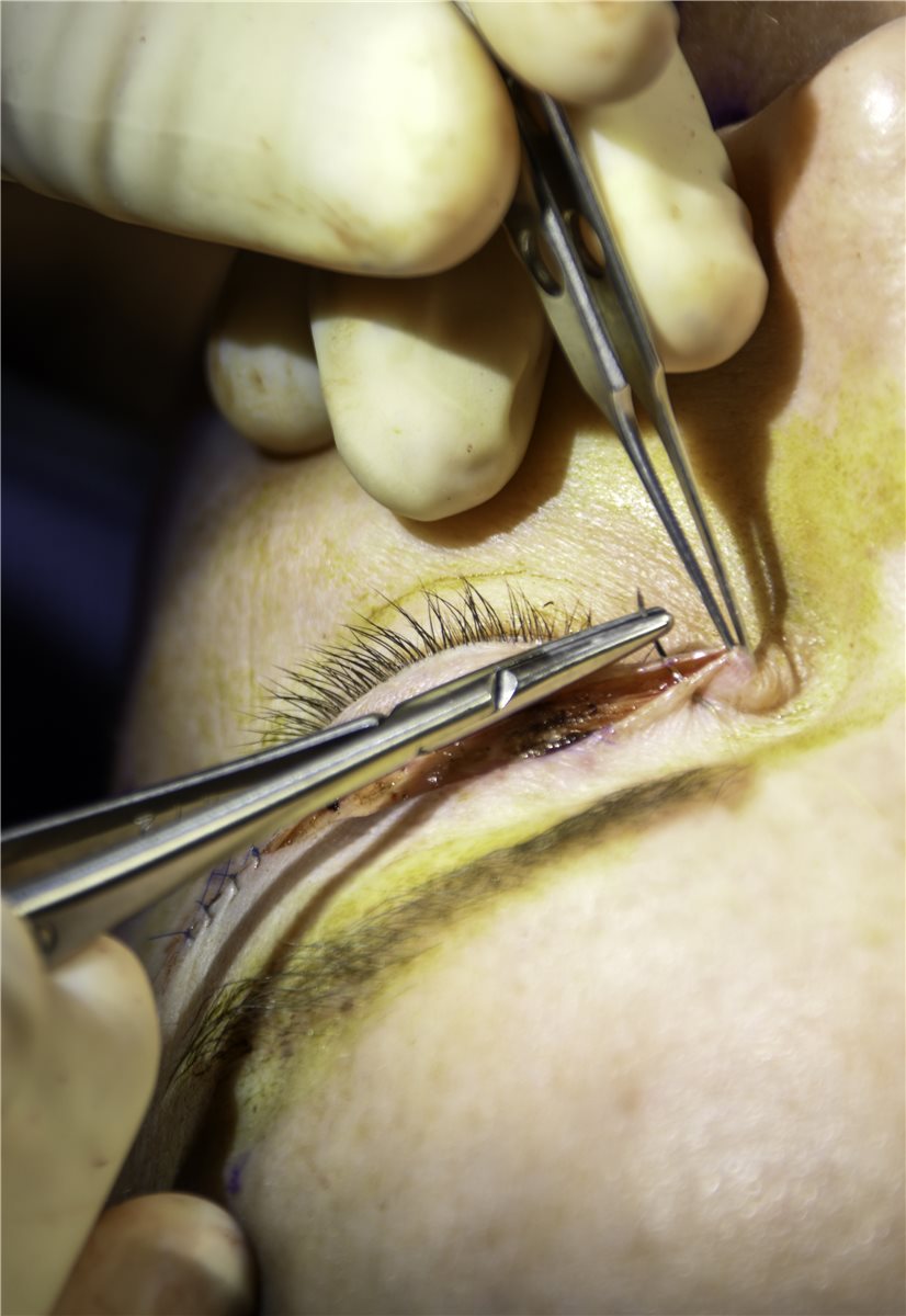
The amount of skin to be excised is marked preoperatively with the patient upright, since lid skin redistributes differently lying down, and marking while supine risks removing too much or too little.
An incision is made along the upper lid crease, and the marked segment of redundant skin, and often a strip of underlying orbicularis muscle, is excised. Prolapsed orbital fat, most often from the medial and central compartments, is conservatively trimmed if contributing to the bulk.
The incision is closed with fine sutures along the natural lid crease, which helps camouflage the resulting scar once healed.
Complications
- Dry eye, often transient, related to temporary changes in blink dynamics and lid closure while swelling resolves; pre-existing dry eye increases this risk and should be addressed before surgery where possible
- Asymmetry between the two sides, which can result from uneven preoperative marking or differential healing, and sometimes needs a minor revision
- Lagophthalmos, incomplete eyelid closure, from excising too much skin; a conservative approach to skin removal is the main safeguard against this
- Haematoma or, rarely, retrobulbar haemorrhage, which is why patients are counselled about warning signs and monitored in the immediate postoperative period
- Scarring or milia along the incision line, generally minor and often resolving or improving without further treatment
Conservative skin excision, erring toward removing slightly less rather than slightly more, is the main technical safeguard against the most functionally significant complication, lagophthalmos, since skin that turns out to be insufficiently removed can always be revised later, while skin that has been over-removed cannot be put back.
Postoperative Course
Swelling and bruising are expected in the first one to two weeks and generally settle with cold compresses and time. Sutures are typically removed within about a week if not self-dissolving.
Final cosmetic and functional result, including any residual visual field improvement, is usually assessed once swelling has fully resolved, generally by around six to eight weeks postoperatively.
Prognosis
Outcomes are generally very good, with high patient satisfaction for both the cosmetic appearance and, where a functional indication was present, measurable improvement in superior visual field.
Recurrence of skin laxity can occur over subsequent years as the natural ageing process continues, though this is a gradual, long-term consideration rather than an early treatment failure, and most patients do not need repeat surgery for many years, if at all.
Concurrent conditions, particularly ptosis or brow ptosis that were not addressed at the same time as the blepharoplasty, remain unresolved regardless of how well the skin excision itself has been performed, which is why accurate preoperative diagnosis of what is actually causing a patient’s appearance matters as much as surgical technique.


Document what you see
Two smartphone imaging tools built for everyday clinic use — one for the slit lamp, one for the fundus.
From Choroida — the team behind this siteReferences
- Damasceno RW, Cariello AJ, Cardoso EB, et al. Upper blepharoplasty with or without resection of the orbicularis oculi muscle. Ophthalmic Plastic and Reconstructive Surgery. 2011.
- American Society of Ophthalmic Plastic and Reconstructive Surgery. Blepharoplasty guidelines.
- Pacella SJ, Nahai FR, Nahai F. Blepharoplasty for the upper eyelid: a systematic approach. Aesthetic Surgery Journal. 2010.
- Upper Eyelid Blepharoplasty. EyeWiki, American Academy of Ophthalmology.
- Blepharoplasty. StatPearls, NCBI Bookshelf.
Test yourself
A few questions straight from this article.
-
Upper eyelid blepharoplasty is performed to correct which underlying eyelid condition?
The procedure removes redundant upper lid skin, and often orbital fat, to correct dermatochalasis — the age-related laxity and excess skin that can hood the lid margin. -
Which change allows orbital fat to prolapse forward and add bulk to the ageing upper eyelid?
Alongside declining collagen and elastin in the skin, the upper lid orbital septum weakens with age, letting orbital fat prolapse forward and contribute to the visible bulk. -
Which test establishes a functional rather than cosmetic indication for upper eyelid blepharoplasty?
Visual field testing with the skin taped up documents how much superior field loss is caused by the skin itself and confirms it improves once the obstruction is lifted. -
Which measurements separate dermatochalasis from a coexisting ptosis before blepharoplasty?
Eyelid position and function are assessed by margin reflex distance and levator function, because ptosis is common in the same age group and must be addressed separately if present. -
Which condition can mimic or worsen the appearance of dermatochalasis and may need treating instead?
A low or descended brow can imitate dermatochalasis; excising eyelid skin alone in such a patient gives an incomplete and sometimes unsatisfying result. -
How is the skin to be excised marked before upper eyelid blepharoplasty?
Lid skin redistributes differently when lying down, so marking is done with the patient upright; marking supine risks removing too much or too little skin. -
What is the main technical safeguard against lagophthalmos after upper eyelid blepharoplasty?
Lagophthalmos follows removing too much skin. Erring toward removing slightly less is the safeguard, since under-resection can be revised later while over-resected skin cannot be replaced. -
Why is the tear film assessed before a patient undergoes upper eyelid blepharoplasty?
Blepharoplasty alters lid closure dynamics and can aggravate existing dry eye, at least temporarily, so the tear film is examined and treated beforehand where possible. -
When is the final cosmetic and functional result of upper eyelid blepharoplasty usually assessed?
Swelling and bruising dominate the first one to two weeks, so the definitive cosmetic result and any residual field improvement are judged once swelling has fully resolved, generally by six to eight weeks. -
What is the expected long-term course after a technically successful upper eyelid blepharoplasty?
Satisfaction is high and field improvement measurable, but ageing continues, so laxity can slowly return; this is a long-term consideration rather than early treatment failure.

Writes and reviews the clinical articles on Educate. Every post is checked against peer-reviewed sources before it goes live.


