
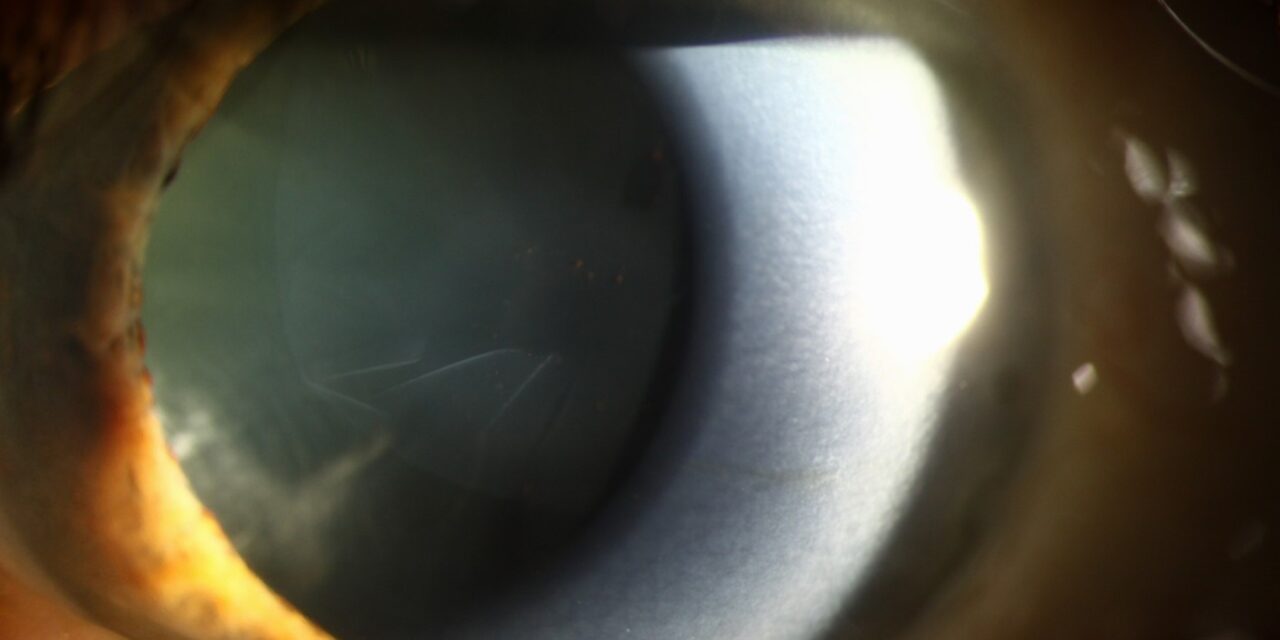
Exfoliation glaucoma is a condition every treating ophthalmologist approaches with a bit more vigilance than typical primary open-angle glaucoma, since this specific secondary glaucoma has a well-documented reputation for higher pressures, more pronounced diurnal pressure fluctuation, and a generally less predictable, sometimes more treatment-resistant course than its more common counterpart.
From Syndrome to Glaucoma
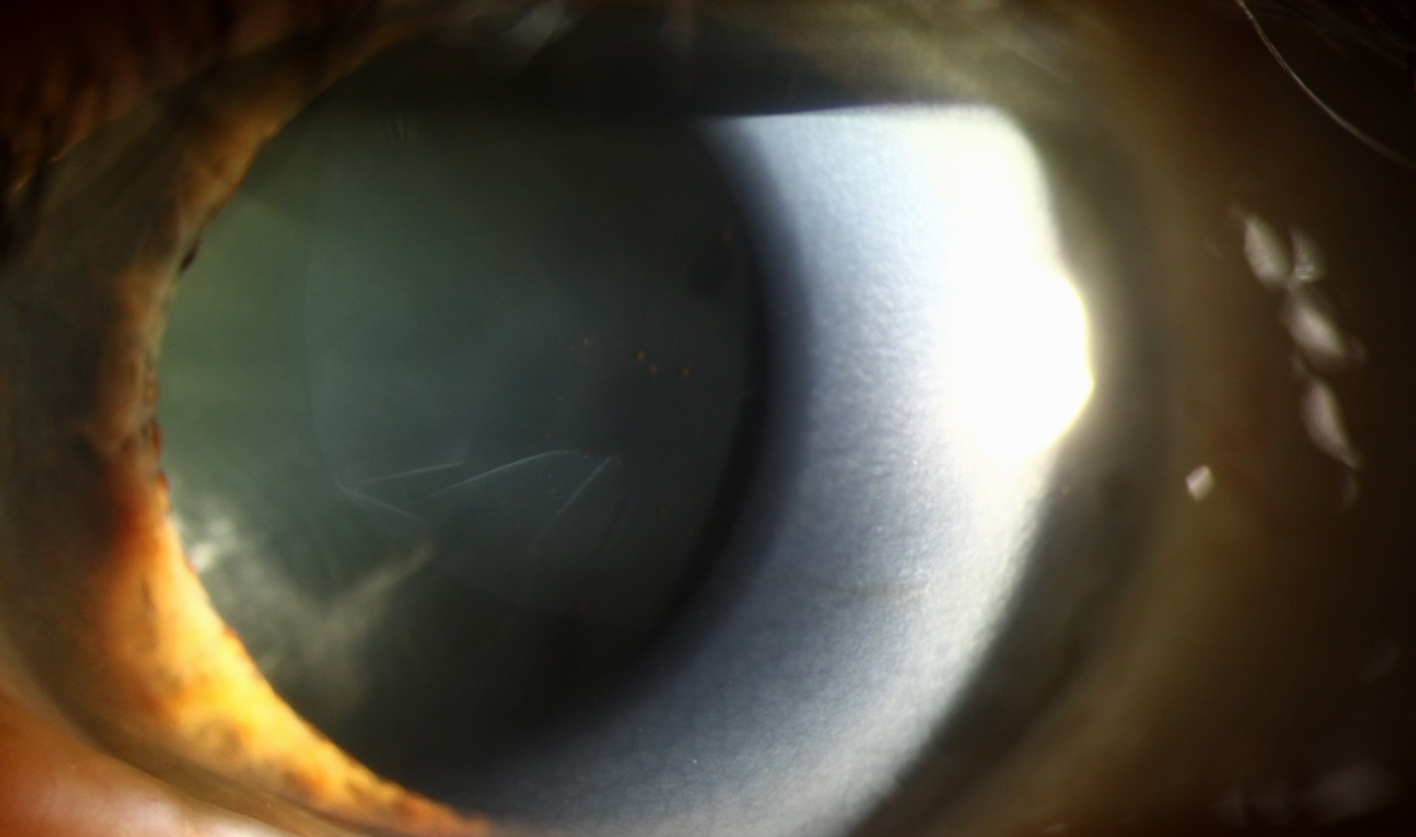
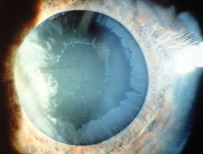
Pseudoexfoliation syndrome, characterized by the deposition of flaky, dandruff-like white material on the lens capsule, pupillary margin, and other anterior segment structures, is common, particularly in older populations, and a substantial proportion of affected patients eventually develop exfoliation glaucoma, related to progressive obstruction of the trabecular meshwork by this same exfoliative material along with associated pigment.
Why Exfoliation Glaucoma Behaves More Aggressively
Several features distinguish exfoliation glaucoma from typical primary open-angle glaucoma in ways that matter directly for management.
- Intraocular pressure at presentation tends to be higher on average, and more patients present with an acute or subacute pressure spike rather than the more gradual, insidious pressure elevation typical of primary open-angle glaucoma
- Diurnal pressure fluctuation tends to be more pronounced, meaning a single office pressure measurement may not adequately capture a given patient’s true peak pressure burden, similar in concept to the exercise-related fluctuation pattern seen in pigmentary glaucoma, though driven by a different underlying mechanism
- The disease often progresses more rapidly and can be more resistant to standard medical therapy compared with primary open-angle glaucoma
- Asymmetry between the two eyes is common, since pseudoexfoliation syndrome itself, and the resulting glaucoma, frequently affects the two eyes unevenly, sometimes substantially so
All-fit Slit-Lamp Adapter
Record and share exactly what you see at the slit lamp. One adapter fits any slit lamp or surgical microscope — and any smartphone.
From Choroida — the team behind this siteClinical Features
- Elevated, often markedly elevated, intraocular pressure, sometimes with documented significant diurnal variation
- Visible pseudoexfoliative material on the anterior lens capsule, classically in a target or bullseye pattern with a central disc, a clear zone, and a peripheral granular zone, along with material often visible at the pupillary margin
- Poor pupillary dilation is a commonly associated finding, related to both the exfoliative material itself and associated iris changes, and this has direct practical implications for cataract surgery planning in these patients
- Optic nerve and visual field changes consistent with glaucomatous damage, assessed using standard techniques
Surgical Considerations Beyond the Glaucoma Itself
Pseudoexfoliation syndrome is also associated with zonular weakness, which carries important implications if the patient requires cataract surgery, given the increased risk of intraoperative complications related to inadequate lens support in these eyes (see zonular dehiscence in cataract surgery and capsular tension ring indications for the related surgical planning considerations this association raises).
Diagnostic Evaluation
- Slit-lamp examination identifying the characteristic pseudoexfoliative material on the lens capsule and pupillary margin
- Gonioscopy, often showing increased, sometimes patchy, trabecular meshwork pigmentation
- Intraocular pressure monitoring, with attention to the possibility of significant diurnal fluctuation not captured by a single measurement
- Optic nerve assessment and visual field testing, following standard glaucoma evaluation principles
- Careful assessment of lens stability (checking for phacodonesis) given the associated zonular weakness risk, particularly important if cataract surgery is anticipated
Management
Medical Therapy
Topical aqueous-suppressant medications are used as initial therapy, though given the tendency toward higher and more variable pressure in this condition, closer monitoring of treatment response and a lower threshold for escalating therapy is often appropriate compared with typical primary open-angle glaucoma management.
Laser Trabeculoplasty
Selective laser trabeculoplasty is often notably effective in exfoliation glaucoma, in some reported series showing a particularly robust initial pressure-lowering response compared with primary open-angle glaucoma, making it an appealing treatment option to consider relatively early in the management sequence for this condition.
Surgical Management
Given the tendency toward more aggressive, sometimes treatment-resistant disease, incisional glaucoma surgery is needed more frequently and sometimes earlier in the course of exfoliation glaucoma than in typical primary open-angle glaucoma, with the specific surgical approach chosen based on disease severity and individual patient factors.
Practical points
Eyes with exfoliation syndrome may have large pressure swings, so a single normal reading does not exclude trouble. Home or diurnal measurements may help. Laser trabeculoplasty is often effective early, although the effect may fade sooner than in other types. Patients should be told that the condition is lifelong, that the other eye may follow, and that cataract surgery needs extra care.
Prognosis
Exfoliation glaucoma generally requires more vigilant, sometimes more aggressive, management than primary open-angle glaucoma, given its tendency toward higher pressure, greater fluctuation, and a less predictable overall course.
With appropriately close monitoring, a lower threshold for treatment escalation, and attention to the specific surgical considerations this condition raises should cataract surgery become necessary, many patients still achieve good long-term pressure control and stable vision, though the more vigilant approach this condition warrants is genuinely important to its favorable management.


Document what you see
Two smartphone imaging tools built for everyday clinic use — one for the slit lamp, one for the fundus.
From Choroida — the team behind this siteReferences
- Ritch R, Schlotzer-Schrehardt U. Exfoliation syndrome. Surv Ophthalmol. 2001;45:265-315.
- Konstas AG, Mantziris DA, Stewart WC. Diurnal intraocular pressure in untreated exfoliation and primary open-angle glaucoma. Arch Ophthalmol. 1997;115:182-185.
- Damji KF, Konstas AG, Liebmann JM, et al. Intraocular pressure following selective laser trabeculoplasty in exfoliation versus primary open angle glaucoma. Br J Ophthalmol. 2006;90:1529-1532.
- Schlotzer-Schrehardt U, Naumann GO. Ocular and systemic pseudoexfoliation syndrome. Am J Ophthalmol. 2006;141:921-937.
