
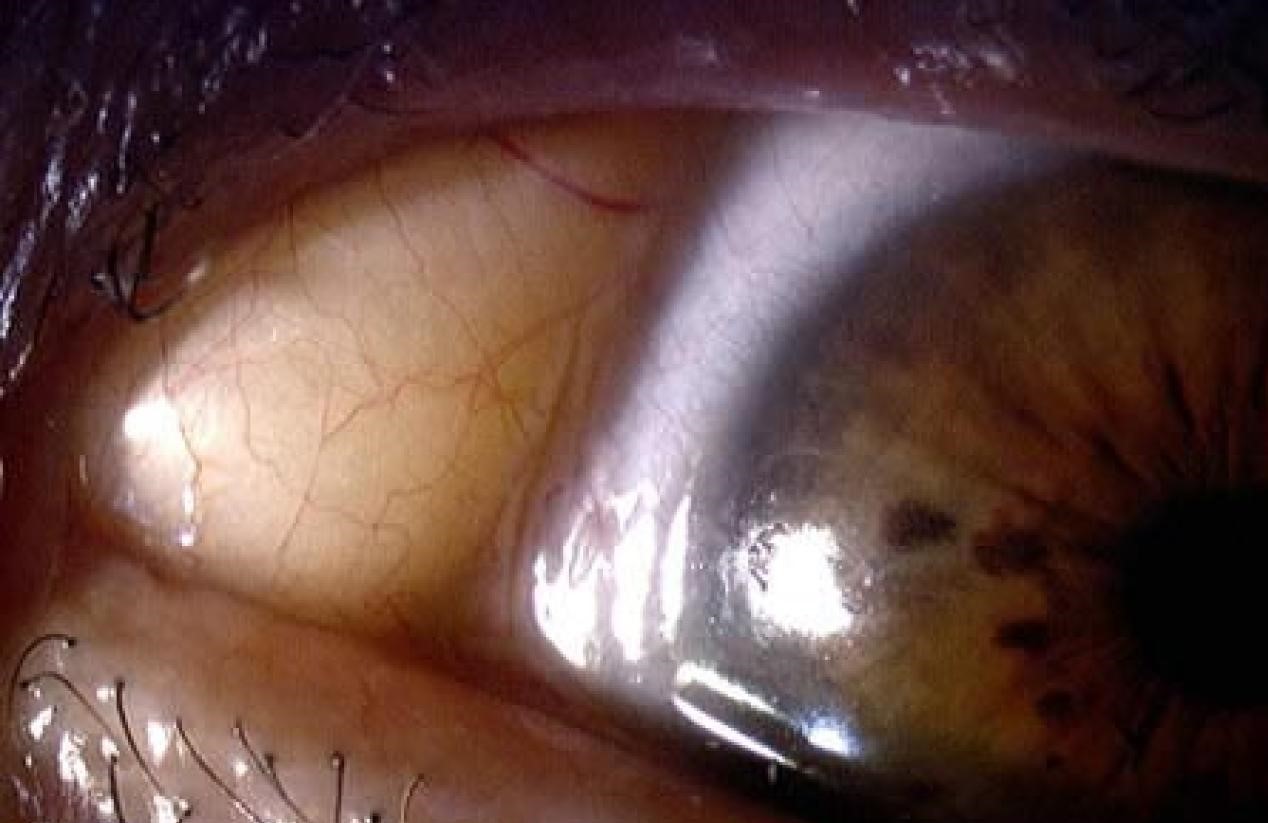
A dermolipoma is a benign congenital choristoma, containing tissue that is normal in composition but abnormal in location, typically presenting as a soft, yellow-white, fatty mass in the superotemporal conjunctiva near the lateral canthus, and recognizing it correctly matters mainly because it is so often benign and stable that overly aggressive surgical attempts at removal cause more harm than the lesion itself ever would.
What a Dermolipoma Is
Like limbal dermoids, dermolipomas are choristomas, congenital lesions arising from tissue displaced during embryonic development rather than acquired growths, and they are composed of a mixture of fibrofatty and, to varying degrees, dermal tissue, which is why they have the characteristic soft, yellowish appearance on examination.
They typically arise in the superotemporal conjunctiva, often extending posteriorly toward or beyond the lateral orbital rim, in contrast to limbal dermoids, which classically sit at or near the corneal limbus itself (see limbal dermoid).
Clinical Presentation
- A soft, mobile, yellow-white mass in the superotemporal bulbar conjunctiva, often noticed by a parent or caregiver, or found incidentally on examination
- Can extend posteriorly into the orbit, sometimes more extensively than is apparent on external examination alone
- Occasionally contains fine hairs, similar to limbal dermoids, given the shared dermal tissue component
- Generally asymptomatic, though larger lesions can occasionally cause mild ocular surface irritation or a foreign body sensation
- Usually does not encroach on the visual axis and typically does not cause significant astigmatism or amblyopia, unlike limbal dermoids, which are more likely to affect the cornea directly given their more central location
All-fit Slit-Lamp Adapter
Record and share exactly what you see at the slit lamp. One adapter fits any slit lamp or surgical microscope — and any smartphone.
From Choroida — the team behind this siteAssociations
Dermolipomas can occur in isolation or as part of Goldenhar syndrome (oculo-auriculo-vertebral spectrum), particularly when bilateral or associated with other findings such as preauricular skin tags, vertebral anomalies, or eyelid coloboma, so recognition of a dermolipoma, particularly in combination with any of these other features, should prompt a broader systemic evaluation for this syndrome.
Diagnosis
Diagnosis is generally made clinically, based on the characteristic appearance and location of the lesion.
Imaging, typically orbital MRI or CT, can be useful when the lesion appears to extend significantly posteriorly, to characterize the full extent of the mass before considering any surgical intervention, and to help distinguish it from other orbital or conjunctival lesions when the presentation is less typical.
Management
Observation
Because dermolipomas are benign, generally stable in size over time, and rarely threaten vision, observation is the appropriate management for the majority of cases, particularly when the lesion is asymptomatic and not cosmetically significant to the patient or family.
When Surgery Is Considered
- Significant cosmetic concern, particularly for larger or more visible lesions
- Ocular surface irritation attributable to the mass
- Rare cases with enough anterior extension to threaten visual function
Surgical Caution
Surgical excision of a dermolipoma requires particular caution, since these lesions often extend posteriorly into orbital fat and can be intimately associated with orbital structures, including the lacrimal gland and extraocular muscles, particularly the lateral rectus.
- Aggressive attempts at complete excision risk significant complications, including damage to the lacrimal gland (which can cause dry eye), extraocular muscle injury (causing diplopia or restrictive strabismus), and ptosis
- For this reason, when surgery is undertaken, a conservative, partial debulking approach, addressing the visible and cosmetically or symptomatically significant anterior portion of the lesion while leaving the deeper, posteriorly extending component undisturbed, is generally preferred over attempting complete excision
Prognosis
Most dermolipomas remain stable and asymptomatic throughout life, requiring no treatment beyond periodic observation.
When surgery is undertaken for cosmetic or symptomatic reasons, a conservative approach generally achieves a good cosmetic result while minimizing the risk of the significant orbital complications that more aggressive excision can cause.


Document what you see
Two smartphone imaging tools built for everyday clinic use — one for the slit lamp, one for the fundus.
From Choroida — the team behind this siteReferences
- Mansour AM, Barber JC, Reinecke RD, Wang FM. Ocular choristomas. Surv Ophthalmol. 1989;33:339-358.
- Shields JA, Shields CL. Eyelid, Conjunctival, and Orbital Tumors: An Atlas and Textbook. 3rd ed. Philadelphia: Wolters Kluwer; 2016.
- Pokharel A, Poudel S, Karki DB. Dermolipoma: a review. Nepal J Ophthalmol. 2013;5:132-135.
- Bansal S, Basu S, Mishra DK. Deep dermolipoma of the orbit. BMJ Case Rep. 2013;2013:bcr2012008337.
