
A staring, wide-eyed appearance is often the first thing anyone notices — and it can appear before the thyroid blood tests do.
Thyroid eye disease (TED), also called Graves’ orbitopathy, is an autoimmune disorder of the orbit and the most common extrathyroidal manifestation of Graves’ disease.
It can occur before, during, or even after a patient’s thyroid hormone levels have been brought under control.
Most cases are mild, but a meaningful minority progress to disfiguring proptosis, double vision, or — rarely — vision-threatening optic nerve compression.
Management has changed substantially in recent years with the arrival of a targeted biologic therapy aimed directly at the disease mechanism.
Recognizing TED early, staging its activity and severity correctly, and knowing when urgent referral is needed are what protect vision in this disease.
What Is Thyroid Eye Disease?
Thyroid eye disease (TED) is an autoimmune inflammatory disorder of the orbital fat and extraocular muscles, most commonly occurring in association with Graves’ disease.
Its hallmark features include:
- Proptosis (forward displacement of the globe) from expansion of orbital fat and muscle
- Upper eyelid retraction, producing the characteristic staring appearance
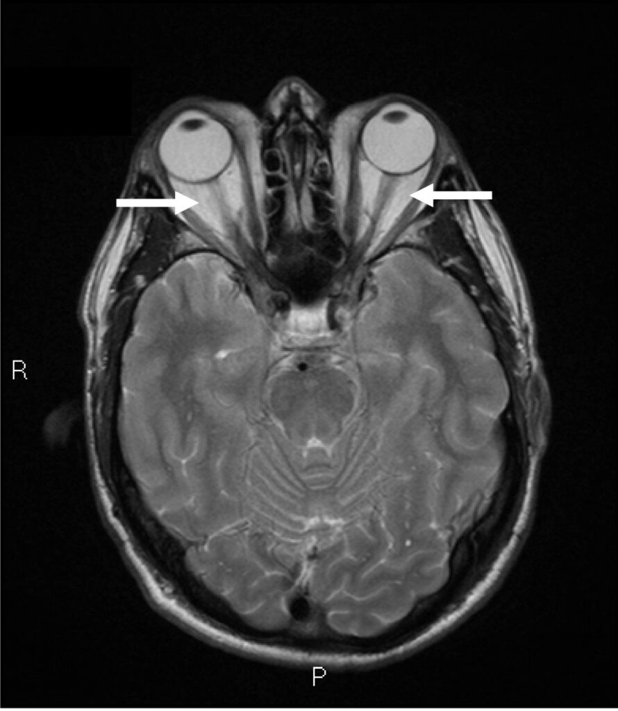
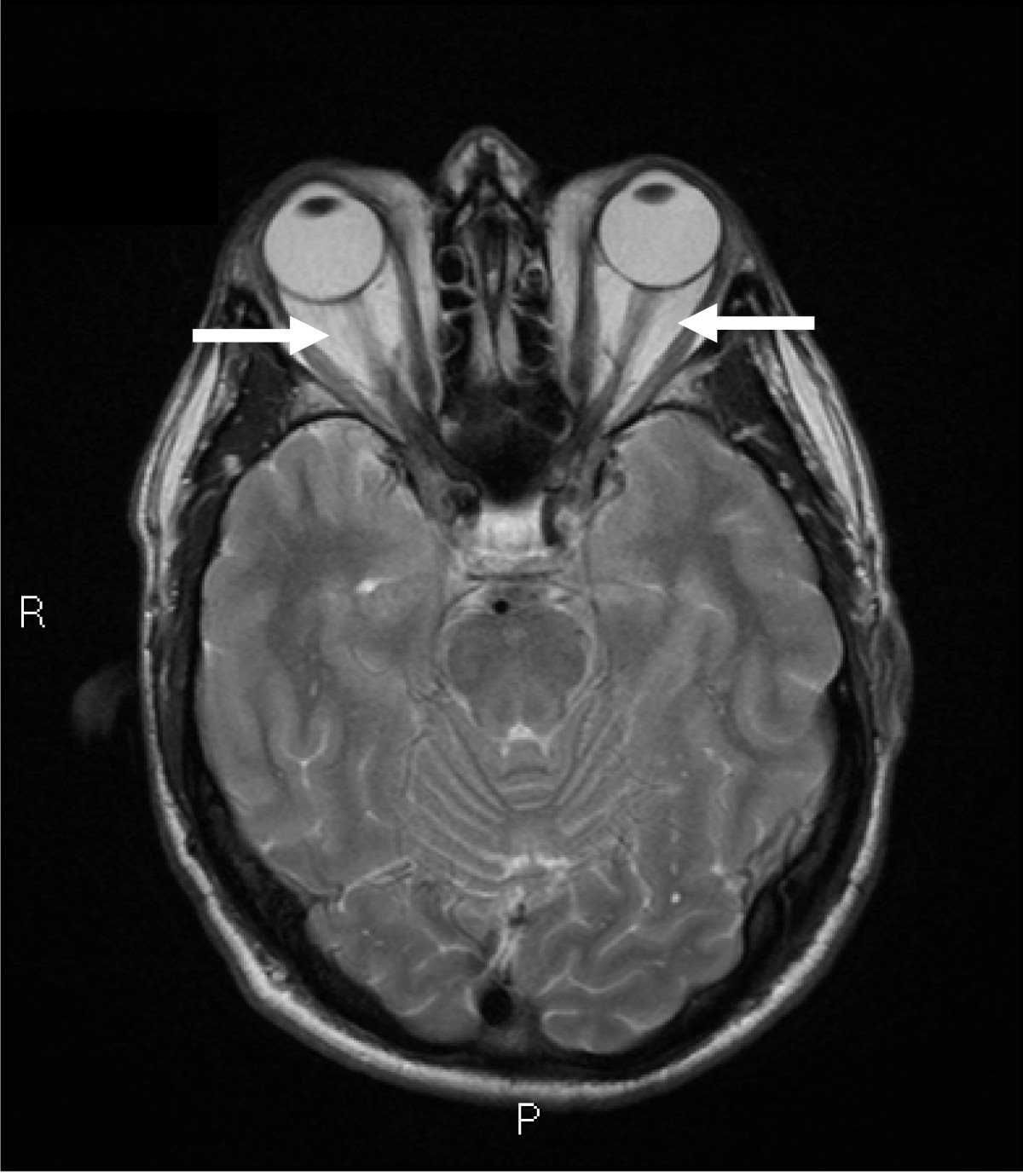
- Extraocular muscle enlargement, which can restrict eye movement and cause double vision
- A spectrum ranging from mild, self-limited disease to severe, sight-threatening involvement
TED severity does not track cleanly with thyroid hormone levels — a euthyroid patient can still have active, progressive orbital disease.
Epidemiology
TED is the most common orbital disease in adults and is closely, though not perfectly, linked to Graves’ disease.
- Most cases occur in patients with hyperthyroid Graves’ disease, though TED can also occur in euthyroid or even hypothyroid patients
- Disease is mild in the majority of patients, moderate-to-severe in roughly a fifth, and rarely sight-threatening in a small minority
- It is substantially more common in women, though men who develop TED often have more severe disease
- Smoking is one of the strongest modifiable risk factors, both for developing TED and for more severe, treatment-resistant disease
Because severity varies so widely, every new diagnosis needs individual staging rather than a one-size-fits-all treatment plan.
All-fit Slit-Lamp Adapter
Record and share exactly what you see at the slit lamp. One adapter fits any slit lamp or surgical microscope — and any smartphone.
From Choroida — the team behind this sitePathophysiology
TED is driven by autoimmune activity directed at antigens shared between the thyroid gland and orbital tissue.
- Autoantibodies against the thyroid-stimulating hormone receptor (TSHR) also act on TSHR expressed in orbital fibroblasts
- Insulin-like growth factor 1 receptor (IGF-1R) on orbital fibroblasts forms a signaling complex with TSHR, amplifying the autoimmune response
- Activated orbital fibroblasts proliferate and produce excess glycosaminoglycans, drawing water into the orbital tissue
- Fibroblasts can also differentiate into adipocytes, expanding orbital fat volume
- The combined effect of muscle enlargement, fat expansion, and edema increases orbital volume within a fixed bony space, producing proptosis and, in severe cases, compressing the optic nerve
This shared antigen mechanism is exactly why treating the thyroid gland alone does not reliably treat the orbital disease driven by the same underlying autoimmunity.
Risk Factors
Strongly Established Risk Factors
- Graves’ disease, the dominant underlying association
- Cigarette smoking — the single strongest modifiable risk factor for both developing and worsening TED
- Radioactive iodine treatment for hyperthyroidism, which can transiently worsen orbital disease in susceptible patients
Other Risk Factors
- Female sex, though male patients often present with more severe disease
- Poor control of thyroid hormone levels, whether hyper- or hypothyroid
- Older age at diagnosis, associated with a higher risk of more severe orbital involvement
Smoking cessation is one of the few interventions a patient can control directly, and it measurably improves both treatment response and long-term outcome.
Clinical Presentation
Symptoms
- A sensation of eye prominence, grittiness, or pressure behind the eyes
- Excessive tearing or dryness from an altered blink and lid position
- Double vision, particularly with restrictive involvement of specific extraocular muscles
- Reduced color vision or visual field loss in the rare cases with optic nerve compression — a genuine emergency
Examination Findings
- Upper eyelid retraction, with visible sclera above the superior limbus (lid lag)
- Proptosis, often measurable and asymmetric between the two eyes
- Restricted extraocular motility, most classically affecting the inferior and medial rectus muscles
- Conjunctival injection and chemosis overlying the insertion of the enlarged rectus muscles
- Relative afferent pupillary defect or reduced color vision in the rare case of optic nerve compression
Any TED patient with reduced vision, color desaturation, or a new visual field defect needs urgent evaluation for optic nerve compression rather than routine follow-up.
Diagnostic Evaluation
Clinical Staging
- Assessment of disease activity (inflammatory signs — redness, swelling, pain) separately from disease severity (proptosis, motility restriction, optic nerve involvement)
- The Clinical Activity Score is commonly used to grade inflammatory activity and guide the choice between anti-inflammatory and rehabilitative treatment
Imaging
- CT or MRI of the orbits to assess extraocular muscle enlargement, orbital fat expansion, and optic nerve crowding at the orbital apex
- Imaging is particularly important when optic nerve compression is suspected or surgical decompression is being planned
Systemic Thyroid Assessment
- Thyroid function tests (TSH, free T4) and TSH-receptor antibody testing to confirm and characterize the underlying thyroid disease
- Coordination with endocrinology, since achieving stable thyroid status supports overall disease control
Distinguishing active, inflamed disease from stable, burnt-out disease is the single most important diagnostic step, since it determines whether anti-inflammatory treatment is likely to help.
Differential Diagnosis
Conditions that can mimic thyroid eye disease include:
- Orbital cellulitis — acute, painful, and typically unilateral, unlike the more gradual, often bilateral course of TED
- Orbital tumors — asymmetric, progressive proptosis without the classic lid retraction and restrictive motility pattern
- Idiopathic orbital inflammation (orbital pseudotumor) — often more acutely painful and typically unilateral
- Carotid-cavernous fistula — pulsatile proptosis with an audible bruit, a distinctive and important mimic to exclude
- Myasthenia gravis — variable, fatigable ptosis and diplopia without the characteristic orbital imaging findings of TED
Bilateral, gradually progressive proptosis with lid retraction in a patient with known or suspected thyroid disease is rarely mistaken for these alternatives once specifically considered.
Management
General Measures
- Smoking cessation, one of the most impactful interventions available
- Optimization and stabilization of thyroid hormone levels in coordination with endocrinology
- Lubrication and ocular surface protection for exposure-related dryness from lid retraction and proptosis
Active, Moderate-to-Severe Disease
- Intravenous glucocorticoids remain a preferred option, particularly when rapid control of inflammation is the priority
- Teprotumumab, an IGF-1R inhibitor, is now established first-line therapy for active, moderate-to-severe disease, particularly when proptosis is prominent, with strong trial evidence for reducing proptosis and diplopia
- Orbital radiotherapy is used in select cases, often alongside glucocorticoids, particularly for restrictive motility disease
Sight-Threatening Disease and Surgical Rehabilitation
- Urgent high-dose glucocorticoids and, where needed, surgical orbital decompression for dysthyroid optic neuropathy
- Staged surgical rehabilitation — orbital decompression, then strabismus surgery, then eyelid surgery — once the disease has become stable and inactive
Surgical rehabilitation is deliberately sequenced and deferred until the inflammatory phase has burned out, since operating on active disease produces unpredictable results.
Prognosis
Most patients follow a self-limited inflammatory course, though the timeline can be long.
- The great majority of cases are mild and improve, though not always fully resolve, once the active inflammatory phase passes
- Moderate-to-severe disease can leave residual proptosis, lid retraction, or diplopia even after inflammation resolves, often requiring rehabilitative surgery
- Dysthyroid optic neuropathy is rare but represents a genuine risk of permanent vision loss if not identified and treated urgently
Long-term outcome depends heavily on early recognition of active disease and prompt treatment during the inflammatory window, before permanent fibrosis sets in.
Would you like to document anterior segment findings with your smartphone?


Document what you see
Two smartphone imaging tools built for everyday clinic use — one for the slit lamp, one for the fundus.
From Choroida — the team behind this siteReferences
- Bartalena L, Kahaly GJ, Baldeschi L, et al. The 2021 European Group on Graves’ Orbitopathy (EUGOGO) clinical practice guidelines. European Journal of Endocrinology. 2021.
- Douglas RS, Kahaly GJ, Patel A, et al. Teprotumumab for the treatment of active thyroid eye disease. New England Journal of Medicine. 2020.
- Comparison of the 2021 EUGOGO guidelines and the 2022 ATA/ETA consensus statement for the management of Graves’ orbitopathy. Journal of Endocrinological Investigation. 2024.
- Bahn RS. Graves’ ophthalmopathy. New England Journal of Medicine. 2010.
- Thyroid Eye Disease. EyeWiki, American Academy of Ophthalmology.
Test yourself
A few questions straight from this article.
-
Thyroid eye disease is the most common extrathyroidal manifestation of which condition?
Thyroid eye disease, also called Graves' orbitopathy, is an autoimmune disorder of the orbit and the most common extrathyroidal manifestation of Graves' disease. -
Which receptor forms a signaling complex with TSHR on orbital fibroblasts in thyroid eye disease?
IGF-1R on orbital fibroblasts complexes with TSHR and amplifies the autoimmune response, which is why it became a therapeutic target. -
What proportion of thyroid eye disease patients have moderate-to-severe disease?
Disease is mild in the majority of patients, moderate-to-severe in roughly a fifth, and rarely sight-threatening in a small minority. -
Which is the single strongest modifiable risk factor in thyroid eye disease?
Smoking is the strongest modifiable risk factor both for developing thyroid eye disease and for more severe, treatment-resistant disease, and cessation measurably improves outcome. -
Which extraocular muscles are most classically involved in restrictive thyroid eye disease?
Restricted motility in thyroid eye disease most classically affects the inferior and medial rectus muscles, with injection and chemosis overlying their insertions. -
Which tool is commonly used to grade inflammatory activity in thyroid eye disease?
Activity is assessed separately from severity, and the Clinical Activity Score guides the choice between anti-inflammatory treatment and rehabilitative surgery. -
Which finding in thyroid eye disease should prompt urgent evaluation for optic nerve compression?
Reduced vision, colour desaturation or a new field defect suggests dysthyroid optic neuropathy, which needs urgent evaluation rather than routine follow-up. -
Which targeted biologic is now established first-line therapy for active, moderate-to-severe thyroid eye disease?
Teprotumumab, an IGF-1R inhibitor, is established first-line therapy for active moderate-to-severe disease, particularly with prominent proptosis, with trial evidence for reducing proptosis and diplopia. -
When should staged surgical rehabilitation for thyroid eye disease be undertaken?
Rehabilitation is sequenced as orbital decompression, then strabismus surgery, then eyelid surgery, and is deferred until inflammation has burned out because operating on active disease gives unpredictable results. -
Which mimic of thyroid eye disease presents with pulsatile proptosis and an audible bruit?
Carotid-cavernous fistula is a distinctive and important mimic to exclude, marked by pulsatile proptosis with an audible bruit.

Writes and reviews the clinical articles on Educate. Every post is checked against peer-reviewed sources before it goes live.


