
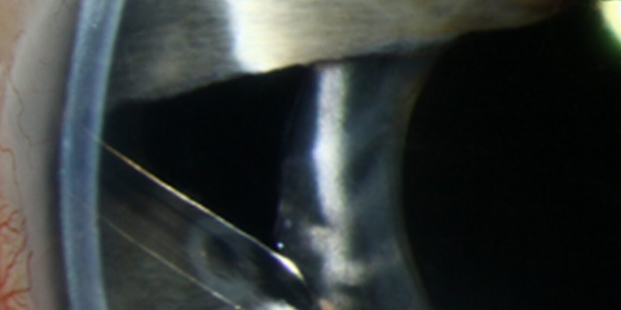
Chandler syndrome is the iridocorneal endothelial syndrome variant in which the cornea, rather than the iris, bears the brunt of the disease, and recognizing this shift in emphasis matters clinically since the corneal edema characteristic of Chandler syndrome can develop and cause visual symptoms even at relatively modest, sometimes near-normal, intraocular pressure readings, a detail that can otherwise make the diagnosis puzzling.
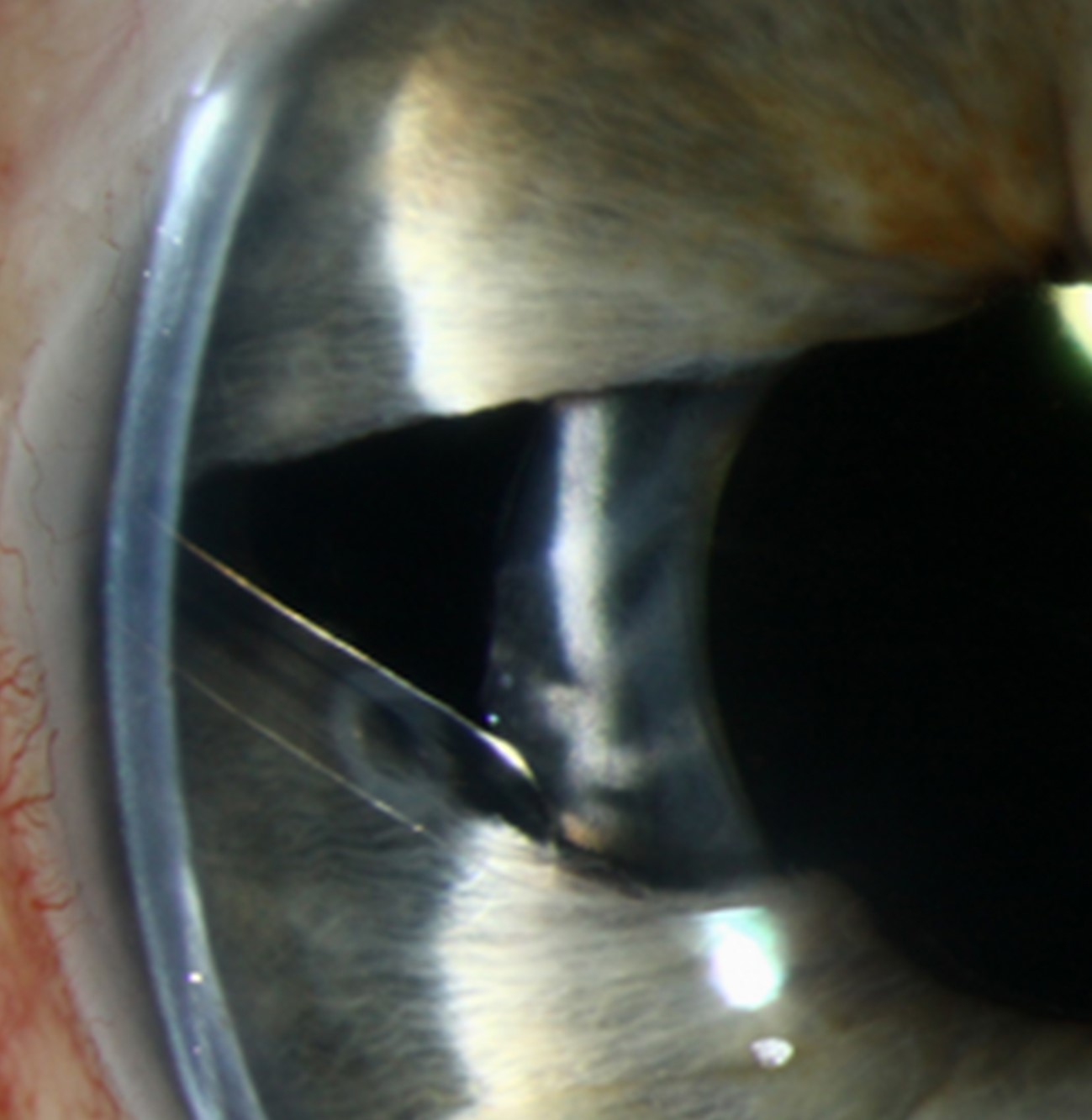
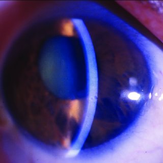
Where Chandler Syndrome Sits in the ICE Spectrum
Chandler syndrome is one of three recognized presentations within the iridocorneal endothelial syndrome spectrum, alongside essential iris atrophy and Cogan-Reese syndrome, all sharing the same underlying mechanism of an abnormal corneal endothelial cell population behaving in an epithelial-like manner, proliferating and migrating across the angle and iris surface (see essential iris atrophy for the more iris-dominant presentation within this same spectrum).
In Chandler syndrome specifically, corneal edema and mild endothelial and angle changes dominate the clinical picture, while iris abnormalities, when present, are typically mild, limited to subtle stromal thinning or minimal corectopia, in clear contrast to the pronounced, progressive iris atrophy and corectopia that define essential iris atrophy.
Why Edema Can Develop at Relatively Normal Pressure
The abnormal endothelial cells in Chandler syndrome function poorly, with reduced pump capacity even in the areas not yet overtly affected by the abnormal membrane, which means the cornea in this condition can be more susceptible to edema at intraocular pressures that a normal, healthy endothelium would tolerate without difficulty.
This is an important diagnostic nuance: corneal edema in a patient with Chandler syndrome does not necessarily indicate markedly elevated pressure, and pressure-focused evaluation alone, without specific attention to the endothelium itself, can miss the underlying mechanism.
All-fit Slit-Lamp Adapter
Record and share exactly what you see at the slit lamp. One adapter fits any slit lamp or surgical microscope — and any smartphone.
From Choroida — the team behind this siteClinical Features
- Corneal edema, sometimes with a fine, hammered-metal or beaten-bronze abnormal endothelial appearance on careful examination, reflecting the same abnormal endothelial cell population seen throughout the ICE syndrome spectrum
- Mild, often subtle iris changes, including fine stromal thinning, generally without the dramatic corectopia or iris hole formation characteristic of essential iris atrophy
- Peripheral anterior synechiae can be present, contributing to secondary glaucoma in some patients, though angle involvement and glaucoma tend to be less severe on average than in essential iris atrophy
- Almost always unilateral, consistent with the broader ICE syndrome pattern
Diagnostic Evaluation
- Slit-lamp examination assessing corneal clarity, the specific abnormal endothelial appearance, and the degree (typically mild) of any iris change
- Specular or confocal microscopy demonstrating the characteristic abnormal, epithelial-like endothelial cell morphology shared across the ICE syndrome spectrum
- Gonioscopy to assess for peripheral anterior synechiae and angle involvement
- Intraocular pressure monitoring, recognizing that edema can occur at relatively modest pressure elevations given the underlying endothelial dysfunction
Management
Managing Corneal Edema
- Hypertonic saline drops and ointment can provide symptomatic relief for mild to moderate edema by helping draw fluid from the cornea
- For more significant, visually limiting edema, endothelial keratoplasty can be considered, replacing the dysfunctional endothelial layer, though as with other ICE syndrome presentations, the ongoing abnormal endothelial cell activity characteristic of this condition can sometimes continue to affect graft tissue over time, an important point for realistic patient counseling before surgery
Managing Intraocular Pressure
When secondary glaucoma is present, management follows standard secondary glaucoma principles, generally beginning with aqueous-suppressant medical therapy, with surgical options, including glaucoma drainage device implantation, considered for inadequately controlled disease.
Monitoring
Given the chronic, slowly evolving nature of Chandler syndrome, ongoing monitoring of corneal clarity, intraocular pressure, and angle status supports early identification of any progression requiring treatment adjustment.
Distinguishing it from Fuchs dystrophy
Chandler syndrome usually presents in a middle-aged woman with unilateral corneal edema, normal or mildly raised pressure, and a mild iris change, while Fuchs dystrophy is bilateral, with guttae in both eyes. Specular microscopy shows pleomorphic, dark-bordered "ICE cells" in the affected eye. Gonioscopy can show peripheral anterior synechiae extending above Schwalbe line, which supports the diagnosis.
Management points
Control any glaucoma, since high pressure worsens edema. Endothelial keratoplasty can restore clarity, but membranes may regrow and recurrence is possible. Counsel the patient about the chronic nature of the condition.
Prognosis
Chandler syndrome generally follows a milder overall course than essential iris atrophy, given its typically less severe iris and angle involvement, though corneal edema can still meaningfully affect vision and, in some patients, eventually requires surgical intervention.
With appropriate management of both corneal edema and any associated secondary glaucoma, many patients maintain reasonable, functional vision over the long term, supported by the sustained monitoring this chronic, unilateral condition warrants.


Document what you see
Two smartphone imaging tools built for everyday clinic use — one for the slit lamp, one for the fundus.
From Choroida — the team behind this siteReferences
- Shields MB. Progressive essential iris atrophy, Chandler’s syndrome, and the iris nevus (Cogan-Reese) syndrome: a spectrum of disease. Surv Ophthalmol. 1979;24:3-20.
- Chandler PA. Atrophy of the stroma of the iris; endothelial dystrophy, corneal edema, and glaucoma. Am J Ophthalmol. 1956;41:607-615.
- Alvarado JA, Underwood JL, Green WR, et al. Detection of endothelial cell membrane antigens in the iridocorneal endothelial (ICE) syndrome. Trans Am Ophthalmol Soc. 1992;90:83-94.
- Sacchetti M, Mantelli F, Rehman A, Bonini S. Successful management of glaucoma associated with iridocorneal endothelial syndrome. BMJ Case Rep. 2013;2013:bcr2012007844.
