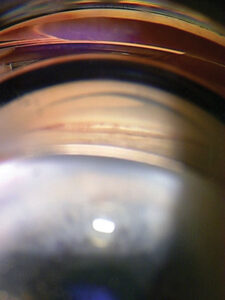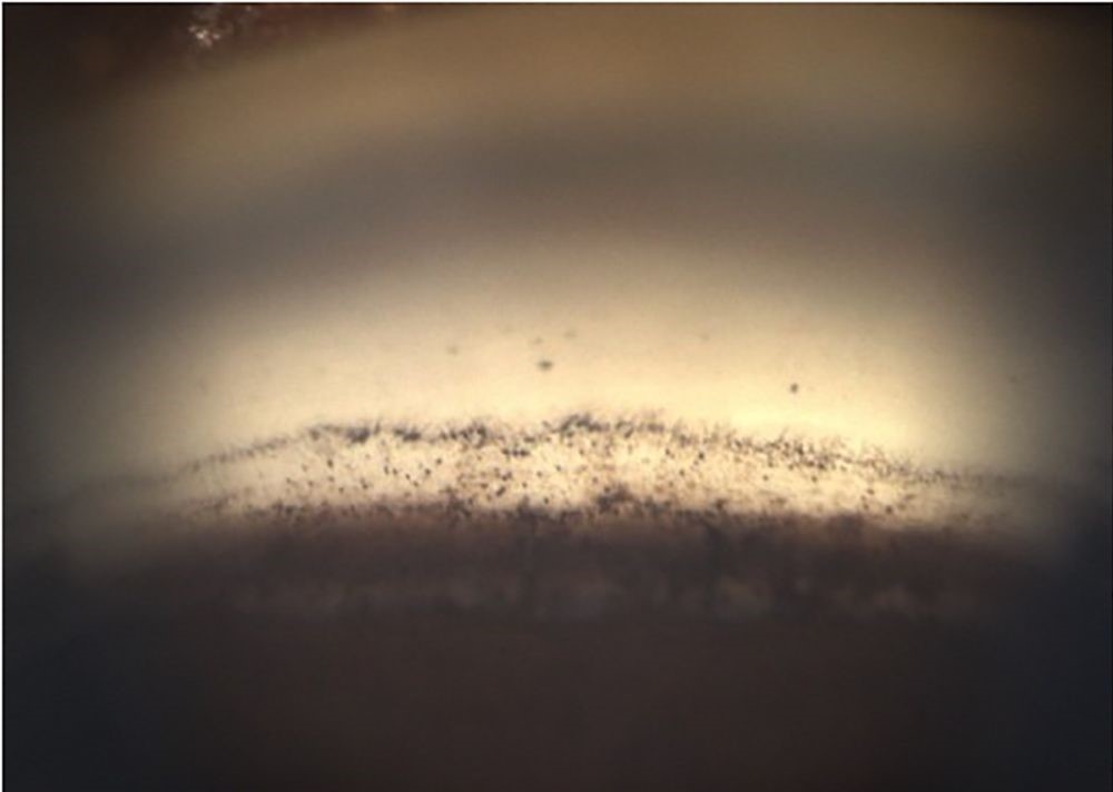
Trabecular meshwork pigmentation is one of the more informative findings available on routine gonioscopy, since the amount and pattern of pigment deposited on the meshwork reflects a history of pigment release within the eye, and the specific degree and distribution of that pigmentation can meaningfully narrow the differential diagnosis for an angle finding that might otherwise be dismissed as simply an incidental variant.
Why Pigment Ends Up on the Trabecular Meshwork
The trabecular meshwork is the primary drainage pathway for aqueous humor, and because of its position and function, it accumulates pigment granules that have been released into the aqueous from the iris pigment epithelium or other pigmented ocular structures, making the degree of trabecular pigmentation, in effect, a visible record of cumulative pigment liberation within that eye over time.
Grading Trabecular Meshwork Pigmentation
Pigmentation is typically graded on a scale, commonly using a system originally described by Scheie, ranging from grade 0 (no visible pigmentation) to grade 4 (dense, heavily pigmented meshwork), providing a semi-quantitative, reproducible way to document this finding and track any change over time.
Comparing pigmentation between the two eyes of the same patient, and correlating the degree of pigmentation with other angle and iris findings, adds further diagnostic context beyond the grade itself.
All-fit Slit-Lamp Adapter
Record and share exactly what you see at the slit lamp. One adapter fits any slit lamp or surgical microscope — and any smartphone.
From Choroida — the team behind this siteCauses of Increased Pigmentation
Pigment Dispersion Syndrome
This is among the most classic causes of significantly increased, often densely homogeneous trabecular meshwork pigmentation, resulting from chronic mechanical rubbing of the posterior iris pigment epithelium against the anterior zonular fibers, releasing pigment that is then carried to and deposited on the trabecular meshwork (see iris transillumination defects for the related mid-peripheral iris finding in this same condition).
Pseudoexfoliation Syndrome
Also commonly associated with increased trabecular pigmentation, though often with a more patchy or irregular distribution pattern than the more homogeneous pigmentation typical of pigment dispersion syndrome, and frequently accompanied by other characteristic findings, including pseudoexfoliative material visible on the anterior lens capsule and pupillary margin.
Prior Intraocular Inflammation
Prior episodes of anterior uveitis or other significant intraocular inflammation can release pigment and inflammatory debris that becomes deposited on the trabecular meshwork, sometimes leaving behind increased pigmentation as a lingering sign even after the active inflammation has resolved.
Prior Trauma
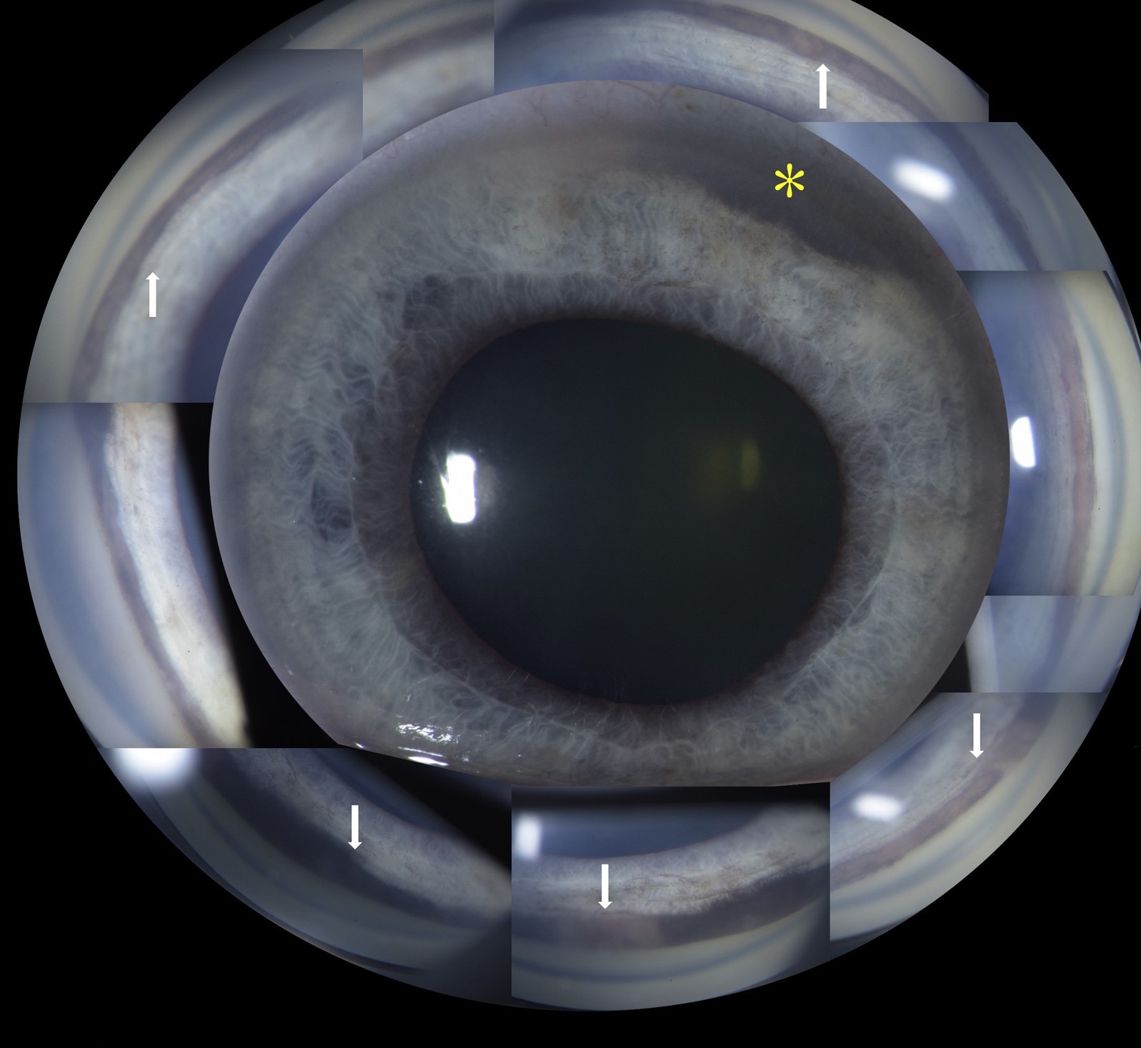
Blunt ocular trauma, including the same mechanism that can produce angle recession, can also release pigment that deposits on the meshwork, sometimes providing an additional supportive clue alongside gonioscopic evidence of angle recession itself (see angle recession glaucoma).
Age-Related Change
Some degree of increased trabecular pigmentation is a recognized, generally benign accompaniment of normal aging, occurring gradually over a patient’s lifetime without necessarily indicating any of the more specific underlying conditions above.
Clinical Significance
- Densely and homogeneously increased pigmentation, particularly in a younger, myopic patient, should prompt specific consideration of pigment dispersion syndrome and assessment for pigmentary glaucoma
- Patchy pigmentation with other supporting findings should prompt consideration of pseudoexfoliation syndrome and its associated glaucoma risk
- Asymmetric pigmentation between the two eyes of the same patient, particularly with a relevant trauma history, supports a traumatic cause, including possible angle recession
- Increased pigmentation is a recognized marker of increased outflow facility variability and, in several of these underlying conditions, is associated with an elevated risk of secondary open-angle glaucoma, supporting closer intraocular pressure monitoring when significant pigmentation is identified
Diagnostic Approach
- Careful, systematic gonioscopy, assessing and grading pigmentation circumferentially around the entire angle, since pigmentation can be asymmetric even within the same eye
- Correlating the pigmentation pattern and degree with other examination findings, including iris transillumination, lens capsule examination for pseudoexfoliative material, and any relevant trauma history
- Intraocular pressure monitoring over time, given the associations between several causes of increased trabecular pigmentation and elevated secondary glaucoma risk
Clinical Value
Trabecular meshwork pigmentation grading, performed as a routine part of gonioscopic examination rather than treated as an incidental afterthought, provides real diagnostic information that, combined with other angle and iris findings, helps identify which patients warrant closer monitoring or further evaluation for an underlying pigment-releasing condition and its associated glaucoma risk.
How to grade the angle
Use a goniolens with low light and a thin beam, examine all four quadrants, and compare the pigment in the posterior trabecular meshwork. Common scales grade pigment from none to dense on a scale of 0 to 4 or 0 to 3. Record the grade in each quadrant, the position of the iris insertion, and the width of the angle. The inferior angle is usually the most pigmented, so compare like with like.
Interpreting the grade
Pigment is more prominent in darker eyes and with age, so a high grade is not abnormal in every case. A dense, uniform band in a young myopic patient suggests pigment dispersion. A patchy, uneven band with a Sampaolesi line suggests exfoliation. In every case, review the pressure and the optic nerve.


Document what you see
Two smartphone imaging tools built for everyday clinic use — one for the slit lamp, one for the fundus.
From Choroida — the team behind this siteReferences
- Scheie HG. Width and pigmentation of the angle of the anterior chamber; a system of grading by gonioscopy. AMA Arch Ophthalmol. 1957;58:510-512.
- Campbell DG. Pigmentary dispersion and glaucoma. A new theory. Arch Ophthalmol. 1979;97:1667-1672.
- Ritch R, Schlotzer-Schrehardt U. Exfoliation syndrome. Surv Ophthalmol. 2001;45:265-315.
- Tesluk GC, Spaeth GL. The occurrence of glaucoma after blunt trauma: a retrospective and prospective study. Ophthalmic Surg. 1985;16:279-285.
Test yourself
A few questions straight from this article.
-
Why does pigment accumulate on the trabecular meshwork?
As the primary drainage pathway for aqueous, the meshwork accumulates pigment granules liberated into the aqueous from the iris pigment epithelium, so its colour records cumulative pigment release in that eye. -
Which practice is recommended when grading trabecular meshwork pigmentation at gonioscopy?
Pigmentation should be assessed and graded circumferentially around the whole angle, recording the grade in each quadrant, because it can be asymmetric even within the same eye. -
What releases pigment onto the trabecular meshwork in pigment dispersion syndrome?
Chronic mechanical rubbing of the posterior iris pigment epithelium against the anterior zonular fibres liberates pigment, which is then carried to and deposited on the trabecular meshwork. -
Which pigmentation pattern is typical of pigment dispersion syndrome?
Pigment dispersion syndrome classically produces significantly increased, often densely homogeneous meshwork pigmentation, and a dense uniform band in a young myopic patient points to this diagnosis. -
Which angle pigmentation pattern suggests pseudoexfoliation syndrome?
Pseudoexfoliation tends to give a more patchy or irregular distribution than pigment dispersion, often with pseudoexfoliative material on the anterior lens capsule and pupillary margin. -
Trabecular pigmentation that is asymmetric between the two eyes, with a relevant history, supports which cause?
Blunt trauma can liberate pigment that deposits on the meshwork, so asymmetric pigmentation with a relevant trauma history supports a traumatic cause and should prompt a search for angle recession. -
How can a previous episode of anterior uveitis affect the trabecular meshwork?
Pigment and inflammatory debris released during anterior uveitis can deposit on the meshwork and leave increased pigmentation as a lingering sign long after the inflammation has settled. -
What is the significance of mildly increased trabecular pigmentation accumulating with age?
Some increase in trabecular pigmentation occurs gradually over a lifetime as a recognised, generally benign age change, without necessarily indicating any specific pigment-releasing disease. -
Which part of the angle is usually the most pigmented?
The inferior angle is usually the most heavily pigmented, so quadrants should be compared like with like when grading and when following a patient over time. -
What does significant trabecular meshwork pigmentation imply for a patient's follow-up?
Several causes of increased pigmentation carry an elevated risk of secondary open-angle glaucoma, so identifying significant pigmentation supports closer intraocular pressure monitoring over time.

Writes and reviews the clinical articles on Educate. Every post is checked against peer-reviewed sources before it goes live.