
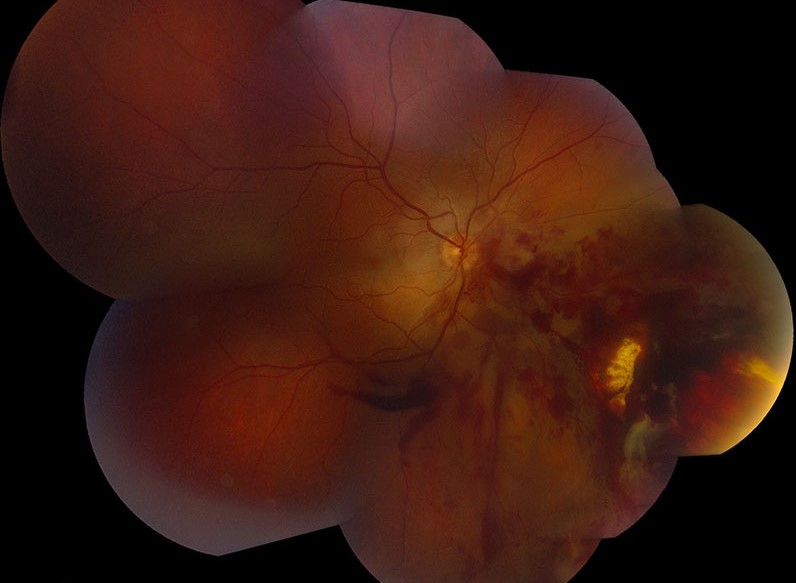
Chorioretinitis sclopetaria is a striking demonstration of how much damage a high-velocity projectile can do to the back of the eye without ever actually penetrating the globe itself, since the injury results from the concussive shockwave of an object passing through the orbit at high speed, close to but not through the eye, rupturing the choroid and retina from the force transmitted through the tissue alone.
The Mechanism: Concussion Without Perforation
Chorioretinitis sclopetaria classically results from a high-velocity missile, most often a bullet or shrapnel fragment, passing through the orbital tissues adjacent to the globe without actually perforating the eye itself, and the sheer force and shockwave transmitted through the periocular and intraocular tissues during this passage is enough to directly rupture the choroid and retina, producing a full-thickness chorioretinal disruption despite the globe technically remaining intact.
Clinical Findings
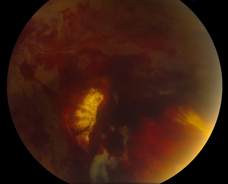
- A large area of chorioretinal disruption and hemorrhage at presentation, often extensive, reflecting the significant concussive force involved
- Over the following weeks to months, the area of disruption evolves and heals through a process of chorioretinal scarring, ultimately producing a well-demarcated, often extensive area of chorioretinal atrophy with variable pigmentation, sometimes described as having a distinctive, somewhat characteristic healed appearance
- A notable and clinically important feature is that, unlike many other severe traumatic chorioretinal injuries, chorioretinitis sclopetaria typically heals through direct adhesion between the retina and underlying tissue at the site of rupture, which generally protects against subsequent retinal detachment at that specific site, in contrast to what might otherwise be feared given the severity of the initial disruption
- Associated findings from the broader orbital and ocular trauma, including vitreous hemorrhage, are common given the significant force involved in the underlying injury
Fundus Explorer Pro
Photograph the retinal findings described here with the phone already in your pocket — 22 D optics and built-in illumination in one handheld unit.
From Choroida — the team behind this siteDistinguishing From Other Traumatic Retinal Findings
- Commotio retinae (Berlin edema) represents retinal whitening from concussive injury without the full-thickness choroidal and retinal disruption characteristic of chorioretinitis sclopetaria, and typically resolves without the permanent, extensive scarring seen in sclopetaria
- A true open-globe injury with retinal involvement involves actual perforation of the globe wall, a fundamentally different mechanism and injury pattern from the concussive, non-perforating mechanism of sclopetaria, with correspondingly different surgical urgency and approach
- Recognizing that the globe has not actually been perforated in chorioretinitis sclopetaria, despite the dramatic degree of posterior segment disruption visible on examination, is an important part of correctly assessing and managing the overall injury
Diagnostic Evaluation
- A history of high-velocity projectile trauma to the orbital or periocular region is central to recognizing this specific mechanism and diagnosis
- Careful examination to confirm globe integrity, since distinguishing a concussive, non-perforating injury from an actual open-globe injury is an essential and urgent early step in any significant orbital or ocular trauma
- Orbital imaging, typically CT, to assess for any retained foreign body, orbital fracture, or other associated injury requiring separate management
- Dilated fundus examination, when the view allows, to characterize the extent of chorioretinal disruption, and B-scan ultrasound when vitreous hemorrhage limits the direct view
Management
Confirming Globe Integrity
The first and most urgent priority in any patient with this injury mechanism is confirming that the globe itself has not been perforated, since this determines the entire subsequent management pathway and urgency.
Supportive Management
Once globe integrity is confirmed, management of chorioretinitis sclopetaria itself is largely observational and supportive, allowing the natural healing process, including the characteristic protective chorioretinal adhesion at the site of injury, to proceed, with monitoring for any complications including delayed retinal detachment, vitreous hemorrhage clearance, or secondary choroidal neovascularization at the margin of the scar.
Managing Associated Injuries
Other aspects of the orbital and ocular trauma, including any orbital fracture, foreign body, or other associated injury, are managed according to their own specific indications, coordinated as part of the overall trauma care.
Prognosis
Despite the dramatic initial appearance of extensive chorioretinal disruption and hemorrhage, chorioretinitis sclopetaria carries a relatively favorable prognosis for retinal attachment specifically, given the protective adhesion that characteristically forms during healing.
Overall visual prognosis depends heavily on whether the macula is involved in the area of chorioretinal disruption, and on the presence and severity of any other associated ocular or orbital injuries sustained in the same traumatic event.


Document what you see
Two smartphone imaging tools built for everyday clinic use — one for the slit lamp, one for the fundus.
From Choroida — the team behind this siteReferences
- Martin DF, Awh CC, McCuen BW 2nd, et al. Treatment and pathogenesis of traumatic chorioretinal rupture (sclopetaria). Am J Ophthalmol. 1994;117:190-200.
- Ahmadabadi MN, Karkhaneh R, Roohipoor R, et al. Traumatic chorioretinitis sclopetaria: clinical features and outcomes. Retina. 2010;30:1279-1284.
- Aizman A, Stein JD, Stenson SM. Chorioretinitis sclopetaria from a paintball injury. Am J Ophthalmol. 2005;140:539-540.
- Sipperley JO, Cunningham CJ, Robertson JE Jr, Machemer R. Traumatic retinopathy in primates: the explanation of commotio retinae. Arch Ophthalmol. 1978;96:2267-2273.
Test yourself
A few questions straight from this article.
-
What mechanism produces the chorioretinal rupture of chorioretinitis sclopetaria?
A bullet or shrapnel fragment travelling through orbital tissue adjacent to the eye transmits enough concussive force to rupture choroid and retina without perforating the globe. -
How does the ruptured area in chorioretinitis sclopetaria typically heal?
Healing occurs through direct chorioretinal adhesion at the rupture site, which generally protects that specific area against subsequent retinal detachment. -
What does the healed lesion of chorioretinitis sclopetaria look like months after injury?
Over weeks to months the disrupted area scars down into an often extensive, sharply bordered zone of chorioretinal atrophy with variable pigment. -
What distinguishes commotio retinae from chorioretinitis sclopetaria?
Commotio retinae is retinal whitening from concussion without full-thickness choroidal and retinal rupture, and it resolves without the permanent extensive scarring of sclopetaria. -
What is the most urgent priority when chorioretinitis sclopetaria is suspected after orbital trauma?
Establishing globe integrity comes first because it determines the entire subsequent management pathway and its urgency. -
Once globe integrity is confirmed, how is the chorioretinal lesion of sclopetaria itself managed?
Management is largely observational, allowing the protective chorioretinal adhesion to form while watching for complications. -
Which test characterises the posterior segment in chorioretinitis sclopetaria when vitreous hemorrhage blocks the view?
When vitreous hemorrhage prevents a direct dilated fundus view, B-scan ultrasound is used to assess the posterior segment. -
Why is orbital CT performed in a patient with chorioretinitis sclopetaria?
CT of the orbit is used to look for a retained foreign body, orbital fracture, or other associated injury needing separate management. -
Which late complication is monitored at the margin of a healed sclopetaria scar?
Follow-up includes watching for secondary choroidal neovascularization arising at the edge of the chorioretinal scar. -
What most strongly determines visual outcome after chorioretinitis sclopetaria?
Overall visual prognosis depends heavily on macular involvement in the zone of chorioretinal disruption, plus any other injuries from the same event.

Writes and reviews the clinical articles on Educate. Every post is checked against peer-reviewed sources before it goes live.


