
Keratoconus posticus is a genuinely important name to parse carefully, since despite sharing “keratoconus” in its title, this rare condition is fundamentally different from classic keratoconus in mechanism, natural history, and prognosis, and confusing the two can lead to unnecessary alarm or inappropriate monitoring for progression that, in the classic form of this rare condition, simply does not occur.
A Different Kind of Curvature Change
Classic keratoconus involves progressive thinning and anterior bulging of the cornea, evolving over years and carrying a real risk of continued ectasia without intervention.
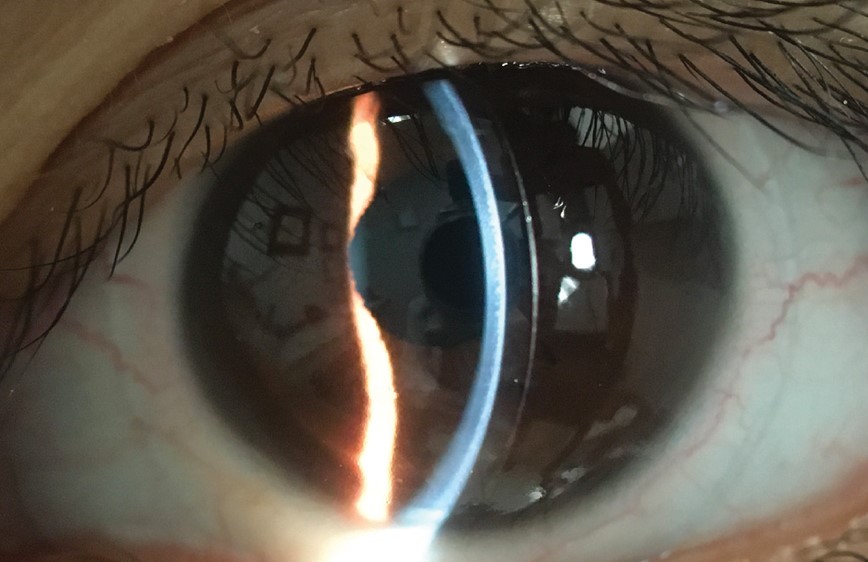
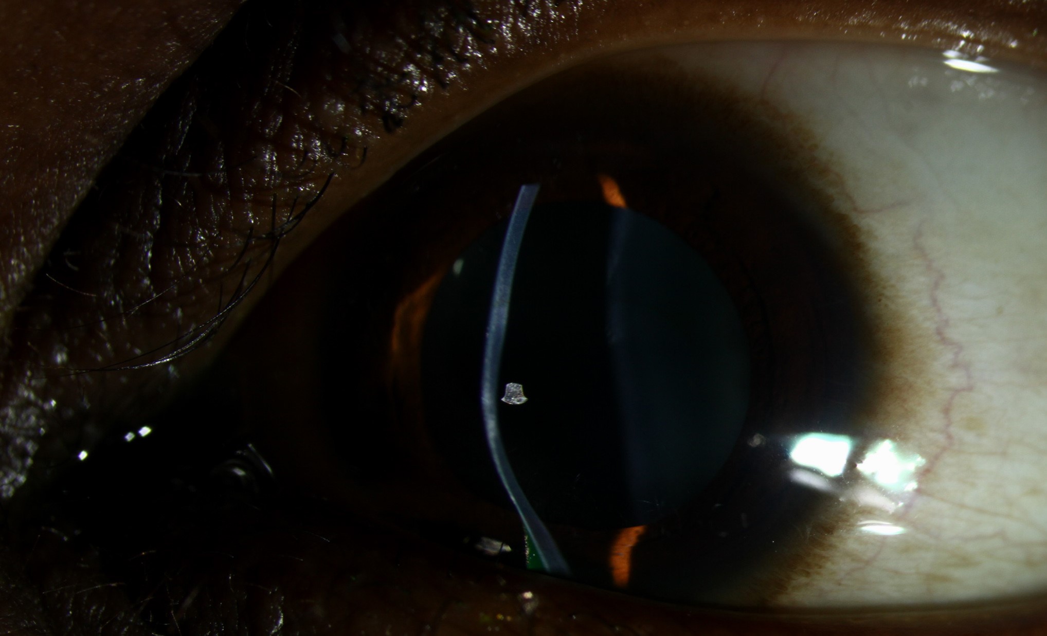
Keratoconus posticus, by contrast, describes an isolated increase in posterior corneal curvature, typically present from birth as a congenital, localized anomaly, and critically, the classic (circumscribed) form is generally non-progressive, representing a stable developmental anomaly rather than an active, evolving ectatic disease process.
Classic (Circumscribed) Keratoconus Posticus
- A localized, often paracentral, area of increased posterior corneal curvature, present from birth
- Generally stable, non-progressive over time, distinguishing it sharply from classic anterior keratoconus in both natural history and long-term implications
- Anterior corneal curvature and thickness are typically normal, or only mildly affected, in contrast to the diffuse anterior thinning and steepening characteristic of classic keratoconus
- Often discovered incidentally on corneal topography or tomography performed for another reason, given the frequent absence of significant symptoms
All-fit Slit-Lamp Adapter
Record and share exactly what you see at the slit lamp. One adapter fits any slit lamp or surgical microscope — and any smartphone.
From Choroida — the team behind this siteGeneralized Keratoconus Posticus
A more diffuse form, generalized keratoconus posticus, has also been described, and this pattern has been associated in some reports with other ocular and systemic developmental anomalies, making it important to distinguish the localized, classically benign circumscribed form from this less common, more diffusely affected pattern when characterizing a given patient’s findings.
Clinical Recognition
- Corneal tomography, imaging both the anterior and posterior corneal surfaces, is essential for correctly identifying keratoconus posticus, since posterior corneal curvature is not reliably assessed by standard keratometry or anterior-surface-only topography
- The key distinguishing feature from classic keratoconus is the combination of an isolated posterior curvature abnormality with a normal or near-normal anterior corneal surface and normal corneal thickness, a pattern not typical of classic anterior ectasia
- Serial tomography over time, demonstrating stability rather than progression, further supports the diagnosis of the classic, benign circumscribed form
Why the Distinction From Classic Keratoconus Matters
- A patient incorrectly diagnosed with classic keratoconus based on an isolated posterior finding might be subjected to unnecessary concern, more frequent monitoring, or even consideration of corneal crosslinking, a treatment aimed specifically at halting the progression that classic circumscribed keratoconus posticus does not exhibit
- Conversely, recognizing a true posterior curvature anomaly supports appropriate reassurance about its generally benign, stable natural history, while still supporting baseline documentation and occasional follow-up imaging to confirm ongoing stability over time, particularly in a patient whose pattern is not unambiguously the classic, well-described circumscribed form
Associated Findings
Keratoconus posticus has been described in association with several other conditions in some reports, including anterior segment developmental anomalies, and a careful, broader anterior segment examination is appropriate when this finding is identified, to assess for any additional developmental features that might warrant further evaluation.
Management
No treatment is needed for the classic, stable, circumscribed form of keratoconus posticus, since it does not progress and generally does not threaten vision significantly on its own, though associated refractive error from the localized curvature change can be corrected with spectacles or contact lenses if visually significant.
Ongoing monitoring with serial tomography is reasonable, particularly when the presentation is not unambiguously classic, to confirm stability over time and distinguish this condition definitively from a true, progressive ectatic process.
Imaging the posterior surface
Slit-lamp examination may show a localized posterior corneal depression with overlying stromal haze, but the diagnosis is confirmed by imaging. Scheimpflug tomography and anterior segment OCT show a steep, localized posterior curvature with relative thinning, while the anterior surface is often normal. Compare both eyes, since the condition may be unilateral, and measure the pachymetry map.
Differential diagnosis and course
Consider Peters anomaly, congenital hereditary endothelial dystrophy, birth trauma, and keratoconus with posterior elevation (see Peters anomaly). Posterior keratoconus is usually congenital, stable, and unilateral, in contrast to keratoconus, which progresses. The main risks to vision are astigmatism and amblyopia in children, which need prompt refraction and treatment.
Prognosis
The classic, circumscribed form of keratoconus posticus carries an excellent, essentially benign prognosis, given its typically stable, non-progressive natural history once correctly identified and distinguished from classic anterior keratoconus.
Correct diagnosis, centered on posterior-surface-inclusive corneal tomography and recognition of the distinct clinical pattern this condition presents, prevents both unnecessary anxiety and unnecessary treatment for a condition that, in its classic form, behaves very differently from the disease its name most readily evokes.


Document what you see
Two smartphone imaging tools built for everyday clinic use — one for the slit lamp, one for the fundus.
From Choroida — the team behind this siteReferences
- Krachmer JH, Feder RS, Belin MW. Keratoconus and related noninflammatory corneal thinning disorders. Surv Ophthalmol. 1984;28:293-322.
- Mannis MJ, Holland EJ, eds. Cornea. 5th ed. Elsevier; 2021.
- Waring GO 3rd, Rodrigues MM, Laibson PR. Corneal dystrophies. II. Endothelial dystrophies. Surv Ophthalmol. 1978;23:147-168.
- Belin MW, Villavicencio OF, Ambrosio RR Jr. Tomographic parameters for the detection of keratoconus: suggestions for screening and treatment parameters. Eye Contact Lens. 2014;40:326-330.
